
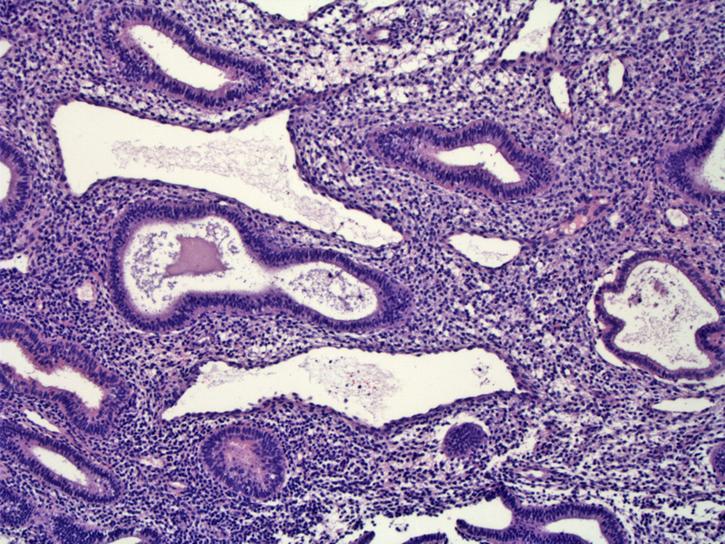
Day 18-19 endometrium: Luminal secretions begin on day 18 and peak at day 20. Note that although luminal secretions are more often found in secretory phase (especially at day 19-20), they are not specific diagnostic features and can be found in proliferative phase endometrium.
)
Day 18-19 endometrium: Note the centrally located nuclei, a feature of day 18 endometrium. Subnuclear or supranuclear vacuoles decrease in size and quantity (compared to day 16 and 17). In this image, the vacuoles are barely visible.
)
Day 19 endometrium: There are scattered subnuclear vacuoles and many of the nuclei are basally located. Note that the endometrium is more orderly, not pseudostratified or mitotic active (compared to day 16 endometrium)
)
In this day 19 endometrium, very few vacuoles are present and the nuclei are basally oriented. The epithelium is not pseudostratified (although tangential sections make it appear otherwise). Note that luminal secretions is prominent in day 19 and peaks at day 20.
)
Day 18 endometrium exhibits both subnuclear and supranuclear vacuoles. With the migration of the subnuclear vacuoles (toward the lumen of the gland), nuclei at the central position is common.
Day 19 endometrium exhibits scattered subnuclear or supranuclear vacuoles, and the nuclei are often at a basal location. There is an increase in luminal secretions. Note that luminal secretions begins on day 18 and peaks at day 20.
Note that day 18-19 endometrium may resemble day 16, however, there are several key features to help you distinguish between the two. In day 16, the endometrium looks "proliferative" with mitotic figures and pseudostratified epithelium. In day 18-19, the endometrium is not mitotically active, and there is only a single (or bilayer) of epithelium without pseudostratification.
1 Mazur MT, Kurman RJ. Diagnosis of Endometrial Biopsies and Curettings. New York, NY: Springer; 2005: 12-19.
2 Nucci MR, Oliva Esther. Gynecologic Pathology: Foundations in Diagnostic Pathology. Philadelphia, PA: Elsevier: 2009: 200.