
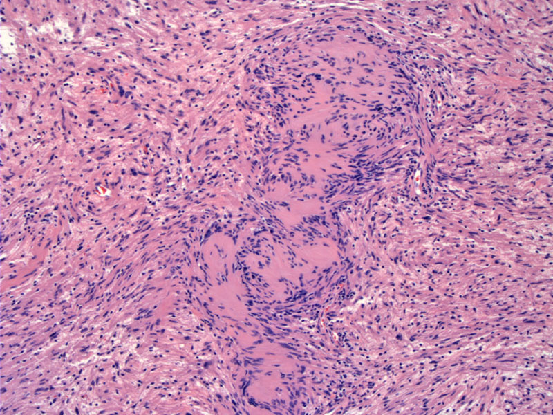
Schwannomas are characterized by Antoni A (spindle cells in a palisading formation known as "Verocay bodies")as seen here, generally admixed with Antoni B (more loosely organized tumor cells that may more cystic spaces) areas. Schwannomas are pseudoencapsulated tumors with a solid tan gross appearance. Bizarre hyperchromatic cells may be seen in ancient schwannomas and should not cause alarm. Blood vessels can be quite prominent. At low power, an Antoni A area is demonstrated with palisading nuclei. Note, however, that palisading nuclei are not specific for schwannomas and can be seen in other entities such as leiomyomas and GISTs.
)
The Verocay bodies can be appreciated here.
)
At even higher power, the neoplastic Schwann cells have fairly bland nuclei without nucleoli. Abundant eosinophilic cytoplasm fills the spindle cells.
)
The looser more edematous regions are designated Antoni B areas.
)
The Schwann cells are tapered spindled cells with slightly wavy nuclei.
)
Tumors or tumor-like conditions arising in peripheral nerves fall into two broad categories: non-neoplastic and neoplastic. The non-neoplastic tumor-like growths include traumatic neuromas, Morton neuromas (a subtype of traumatic neuroma) and palisaded encapsulated neuromas. Neoplastic benign growths include benign tumors such as schwannomas, neurofibromas and perineuriomas. The malignant tumors are grouped under the term "malignant peripheral nerve sheat tumors" (Rosai).
Schwannoma (a.k.a neurolemoma) is an encapsulated profileration of Schwann cells. The mass is actually located outside of the nerve, although at the periphery of the tumor, nerve fibers can be identified.
The tumor cells are positive for S-100, calcineurin, basal lamina components (e.g. laminin), and vimentin, among others (Rosai). Multiple schwannomas and bilateral acoustic schwannomas are indicative of Neurofibromatosis type 2.
Frequently asymptomatic and detected incidentally, although some produce compression symptoms, particularly in children.
Largely a benign tumor. Malignant degeneration is exceptionally rare.
Rosai, J. Rosai and Ackerman's Surgical Pathology. 9th Ed. Philadelphia, PA: Elsevier; 2004: 2264-5.