
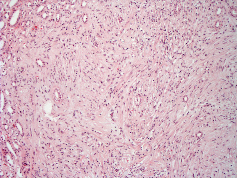
Renomedullary interstitial cell tumors consists of a proliferation of small stellate or polygonal cells arranged in poorly formed fascicles. Entrapped medullary tubules are often found within the lesion, especially at the tumor periphery.
Bland stellate or spindled cells are embedded in a loose hypocellular stroma. Though not demonstrated here, some lesions may be hyalinized or contain deposits of amyloid.
Renomedullary interstitial cell tumor is an extremely common benign entity located in the medulla of the kidney. In fact, these white-gray nodules are found in up to 50% of kidneys in autopsy studies.1,2 These tumors arise from interstitial cells of the renal medulla. Although the function of interstitial cells involves the regulation of blood pressure, these tumors do NOT cause hypertension, and are in fact, completely asymptomatic.
These tumors are asymptomatic and are found incidentally. Grossly, the lesion is a grey-white nodule (1-5 mm) located in the medullary pyramid. These tumors are usually found in individuals over 18 years old.
Excellent; completely benign entity.
1 Zhou M, Magi-Galluzzi, C. Genitourinary Pathology: Foundations in Diagnostic Pathology. Philadelphia, PA: Elvesier; 2006: 329-330.
2 Murphy WM, Grignon DJ, Perlman EJ. Tumors of the Kidney, Bladder and Related Urinary Structures. AFIP Atlas of Tumor Pathology. Fourth series, Fascicle 1. Washington DC; AFIP: 2004; 194-5.

)
)