System: Hematopathology: Lymph Nodes: Benign: Vascular Transformation of Sinuses

System: Hematopathology: Lymph Nodes: Benign: Vascular Transformation of Sinuses

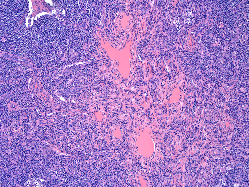
The sinuses are replaced by a vascular proliferation fillfilled with lymph-like fluid. Other examples may show empty lumens, or lumnes congested with blood, or occasionally thrombosed. Image
An angiomatous proliferation is seen surrounded by eosinophilic smooth muscle cells. The more cellular forms may be mistaken for Kaposi's sarcoma, but can be distinguished by the pure sinusoidal distribution, a lack of well-formed spindle cell fascicles, associated fibrosis, and the failure to involve the capsule itself, which is frequently affected by Kaposi's sarcoma Image
This example contains more red cells making the diagnosis a bit less sneaky. Image
The vascular channels lack atypia. Image
)
)
)
)