System: Gynecological: Cervix: Pre-Neoplastic: Keratinizing Dysplasia

System: Gynecological: Cervix: Pre-Neoplastic: Keratinizing Dysplasia

Case 1: Inflamed cervix lined by squamous epithelium producing surface keratin.
Gland is colonized by dysplastic squamous epithelium that appears attenuated, with abundant keratin filling the lumen.
Surface dysplasia is present, which extends into the keratinized layer.
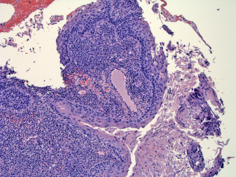
Case 2: Keratinized dysplasia overlies obvious CIN III epithelium.
In the accompanying endocervical curettage, fragments of atypical keratinized squamous epithelium is present.
Keratinizing dysplasia can overlie mildly dysplastic epithelium (CIN I) or moderately or severely dysplastic epithelium (CIN II/III). However, if single or a few dysplastic keratinized squames are seen on a Pap smear, you could not predict whether these cells overlie a CIN I, II or III. Potentially, atypical squames could also be present in inflammed epithelia as well.
)
)
)
)
)