System: Gastrointestinal: Small Intestines: Inflammatory: Crohn's Disease

System: Gastrointestinal: Small Intestines: Inflammatory: Crohn's Disease

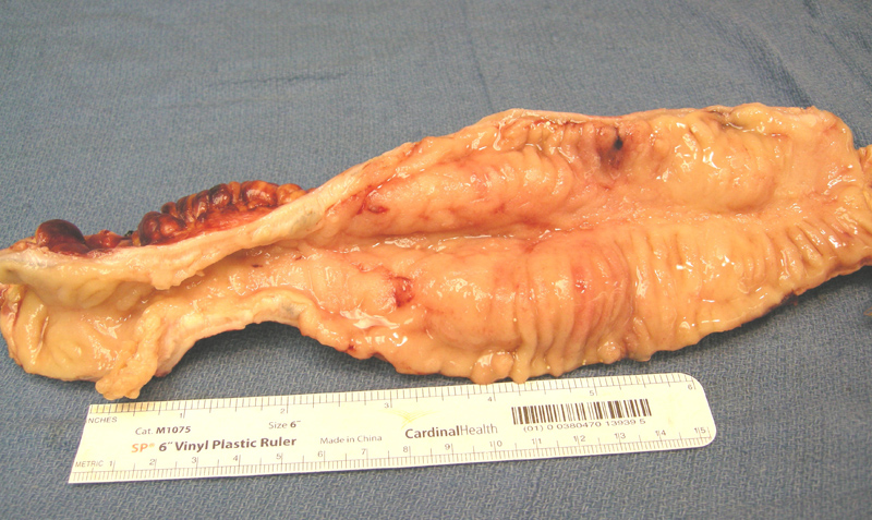
Thickening of the wall and a distinct stricture are seen here. An elevated cancer risk is seen in strictured areas which therefore require close monitoring.
Nuetrophils and a capillary proliration make up the flattened base of this aphthous ulcer.
An aphthous ulcer is progressive into a fissure. Aphthous ulcers often form with adjacent relatively normal mucosa. A neutrophilic infiltrate aggregates at the base of the ulcer leading to deeper penetration into the wall.
Transmural inflammation is seen here, which is a feature of Crohn's and is lacking in ulcerative colitis. Lymphoid aggregates, with or without germinal centers, may be seen in the bowel wall.
This segment of small intenstines shows severe architectural disturbance. The normally regularly spaced villous processes are blunted and irregular, making it difficult to even discern that this is enteric mucosa.
These altered villous projections show mucin depletion and abnormal serrations of glandular outlines.
A pseudopolyp is seen here as a small projection of mucosa covered with granulation tissue and a fibrinous cap.
An intramucosal granuloma contains epithleioid cells loosely aggreated together immediately beneath the surface epithelium. They may be seen in involved as well as uninvolved areas of the bowel, at any level within the bowel wall.
Some granulomas are more of a loose micro-aggregate of epithelioid histiocytes.
The ileal mucosa shows marked acute ileitis with a neutrophilic infiltrate and a crypt abscess along with a granuloma. The architecture is that of a colonic morphology.
This nicely formed epithelioid granuloma parts the gastric glands in the antrum.
Architecturally abnormal villi and hypermucinous epithelium are present. Image
Again, the villi are dysmorphic albeit subtle. Image
Crohn disease is an chronic inflammatory process which can affect any level of the GI tract. The most commonly affected site is the ileum, however, the colon, duodenum, stomach, esophagus and the mouth can be involved (Iacobuzio). Typical manifestations include discontinuous involvement of different segments of the GI tract, transmural inflammation, and complications such as strictures, abscesses, and fistulae.
Current evidence suggests a disruption of mucosal immune homeostasis in genetically susceptible individuals, resulting in altered processing of enteric antigens, pathogenic T cell activation, and chronic inflammation. The disease has a predilection for Western countries (especially Northern Europe) and there is genetic susceptibility component. In the US, people of Jewish descent are 3-5 times more likely to be affected (Kumar).
Endoscopy shows deep, linear ulcerations that may occur as segmental areas of mucosal involvement separated by areas of normal intervening mucosa (“skip lesions”). The rectum is often spared. The mucosa may have a cobblestone appearance due to alternating areas of depressed ulcers and edematous mucosa.
Histologic features include transmural inflammation, glandular effacement, mucosal erosions, cryptitis (collections of neutrophils within the crypts). Noncaseating granulomas can be seen in about half the cases. There is variable inflammation not only grossly, but also microscopically, meaning an inflamed gland can be situated next to a completely normal gland.
Most patients present in the 2nd decade of life but there is a 2nd peak in the 50s-60s. Clinical features depend on its localization and frequently include diarrhea, abdominal pain, fever, clinical signs of subileus or ileus, and/or the passage of blood and mucus per rectum. Fissures can develop and penetrate the bowel wall, leading to serositis, adhesions and formation of fistula tracts between the GI tract and vagina (enterovaginal), bladder (enterovesical) or skin (enterocutaneous).
Extra-intestinal manifestations may also arise and affect the bones and joints, skin, eyes, hepatobiliary system, lungs, and kidneys. They can occur prior to, in conjunction with, or subsequent to active bowel disease. Some specific more common manifestations include erythema nodosum, pyoderma gangrenosum, uveitis, osteoporosis, spondyloarthropathies and primary sclerosing cholangitis.
Medical therapy includes aminosalicylates (5-aminosalicylic acid [5-ASA] agents), corticosteroids, immunosuppressive agents, antibiotics, and biologic agents. The only biologic therapy approved by the FDA is infliximab, an antibody against tumor necrosis factor-α. Refractory fistulas may require surgical resection. In general with Crohn's, surgical resection is onl a palliative option.
Routine surveillance colonoscopy is advocated for detection of dysplasia and malignancy. For those in whom a dysplastic lesions or early malignancy are identified, colectomy can be a potentially curative procedure for both the cancer and the colitis.
Dysplasia and adenocarcinoma arise with increased frequency, and are more likely in those who developed CD at a young age or have a greater extent or severity of disease. Coexisting primary sclerosing cholangitis also puts one at risk. One of the serious complications is the potential development of toxic megacolon.
→Crohn disease is a non-continguous chronic inflammatory disease affecting any segment of the bowel wall and shows transnmural involvement
→Fissures and strictures are common complications.
→Granulomas may be seen in about half the cases.
Iacobuzio-Donahue CA, Montgomery EA. Gastrointestinal and Liver Pathology: Foundations in Diagnostic Pathology. Philadelphia, PA: Elsevier; 2005: 168-172.
Kumar V, Abbas AK, Fausto N. Robbins and Cotran Pathologic Basis of Disease. 7th Ed. Philadelphia, PA: Elsevier; 2005: 847-8.
)
)
)
)
)
)
)
)
)
)
)
)
)