
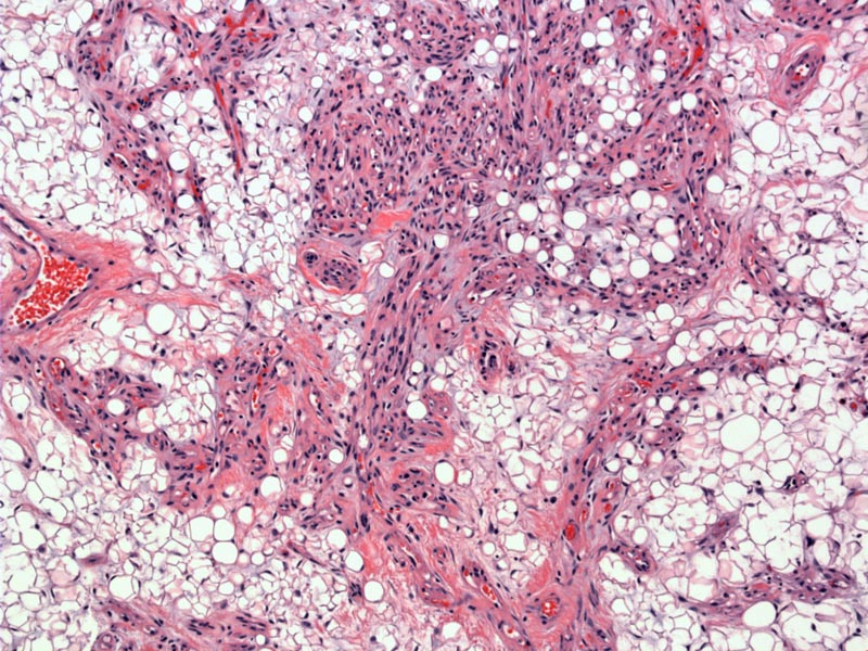
Angiolipomas are very similar to lipomas, but with the added component of small thin-walled blood vessels. The vascular component can range from minimal to over 90% of the tumor, thus, giving the tumor a spindled cellular look.
)
Although not especially prominent in this image, microthrombi within the capillaries is often a helpful diagnostic feature. Note also that not only do endothelial cells proliferate, but the pericapillary pericytes as well.
)
Prominent microthrombi are appreciated a different case of angiolipoma. Look for capillary lumens that have been filled with deep pink material.
)
Another area that is less cellular but illustrates another microthrombus.
)
Predominantly affects males and presents in early adulthood. Multiple lesions are common, and forearm and trunk are frequently affected. The lesions are painful and tender.
Excision.
Excellent. No recurrence after excision, however, new lesions elsewhere may develop.
1 Fletcher CDM, ed. Diagnostic Histopathology of Tumors. 3rd Ed. Philadelphia, PA: Elsevier; 2007:1531.