
This is a case of embryonal rhabdomyosarcoma arising from the prostate of a 15 year old boy. The classic histology consists of small, round undifferentiated cells embedded in a myxoid stroma.
)
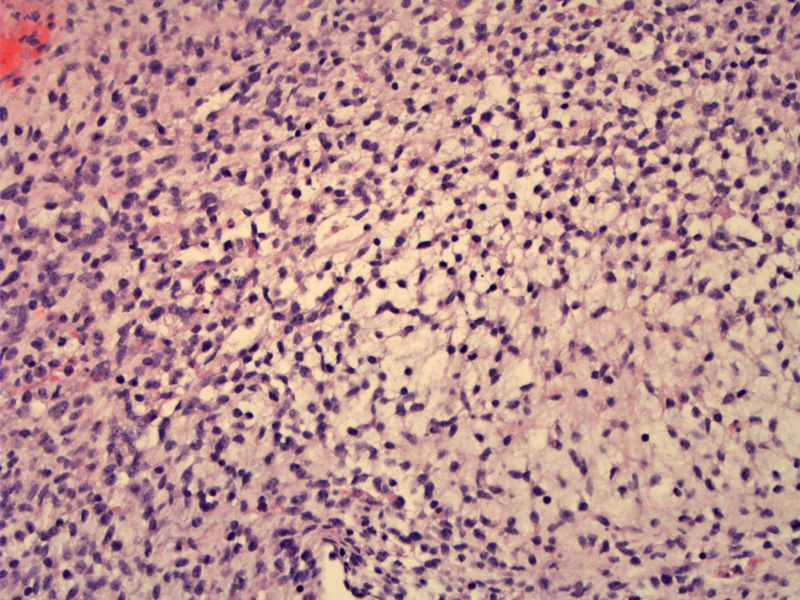
The presence of cellular areas (usually surrounding blood vessels) alternating with paucicellular regions is highly characteristic of embryonal rhabdomyosarcoma.
)
Again, a less cellular area with abundant myxoid stroma with areas of increased cellularity. The tumor cells have scant cytoplasm.
)
One can appreciate cross striations in these strap cells (rhabdomyoblasts).
)
Embryonal rhabdomyosarcoma accounts for 60% of childhood cases of rhabdomyosarcomas. These tumors typically occur in children under 10 years old and arise in the nasal cavity, orbit, middle ear, and urogenital regions.1,2
The classic histology consists of small, round or spindled undifferentiated (basophilic) cells embedded in a myxoid stroma. Note that these cells are smaller than those found in the alveolar subtype. A variable number of rhabdomyoblasts are also seen - cytoplastic cross-striations are only seen in up to 1/3 of these rhabdomyoblasts.1
Botryoid rhabdomyosarcoma (botryoides = bunch of grapes in Greek) is a subset of embryonal rhabdomyosarcoma that arise beneath mucosal surfaces. Also known as sarcoma botryoides, these tumors are distinguished by a subepithelial cambium layer of condensed undifferentiated tumor cells underneath the mucosal surface. They grow in a exophytic, grape-like fashion and protrude into hollow structures such as bladder, vagina and nasopharynx.1,2
In contrast to alveolar rhabdomyosarcoma which exhibit a particular translocation, notably t(2;13)(q35;14) or t(1;13)(q36;q14) which forms the PAX3-FKHR or PAX7-FKHR fusion gene, the major genetic aberration in embryonal rhabdomyosarcomas consists of a deletion in the short arm of chromosome 11 (an allelic loss of 11p15.5).
IHC studies demonstrate that the tumor cells are positive for desmin, MyoD1 and myogenin (myf4).
Most cases arise in children between ages 3-12 years (but can also be seen in younger children and adults). In contrast, alveolar rhabdomyosarcomas tend to affect older age group, between 10-25 years. The tumors tend to occur in the head and neck region, especially orbit, nasopharynx, oral cavity or middle ear. Other common anatomic locations include the retroperitoneum, bile ducts and urogenital tract. Again in contrast, alveolar rhabdomyosarcomas tend to arise in the extremities.
Grossly, an embryonal rhabdomyosarcoma is poorly delineated, soft and white. The botryoid subtype grow beneath the mucosal surface of a hollow organ (like the vagina) and protrude like a bunch of grapes.3
Embryonal rhabdomyosarcomas generally have a more favorable outcome than alveolar rhabdomyosarcomas. With surgery and multidrug therapy, over 80% of children have long-term survival when disease is localized.3
• Myogenic : Rhabdomyosarcoma, Alveolar type
1 Fletcher CDM, ed. Diagnostic Histopathology of Tumors. 3rd Ed. Philadelphia, PA: Elsevier; 2007: 1567-1570.
2 Kumar V, Abbas AK, Fausto N. Robbins and Cotran Pathologic Basis of Disease. 7th Ed. Philadelphia, PA: Elsevier; 2005: 1321-2.
3 Rosai, J. Rosai and Ackerman's Surgical Pathology. 9th Ed. Philadelphia, PA: Elsevier; 2004: 2301-4.