
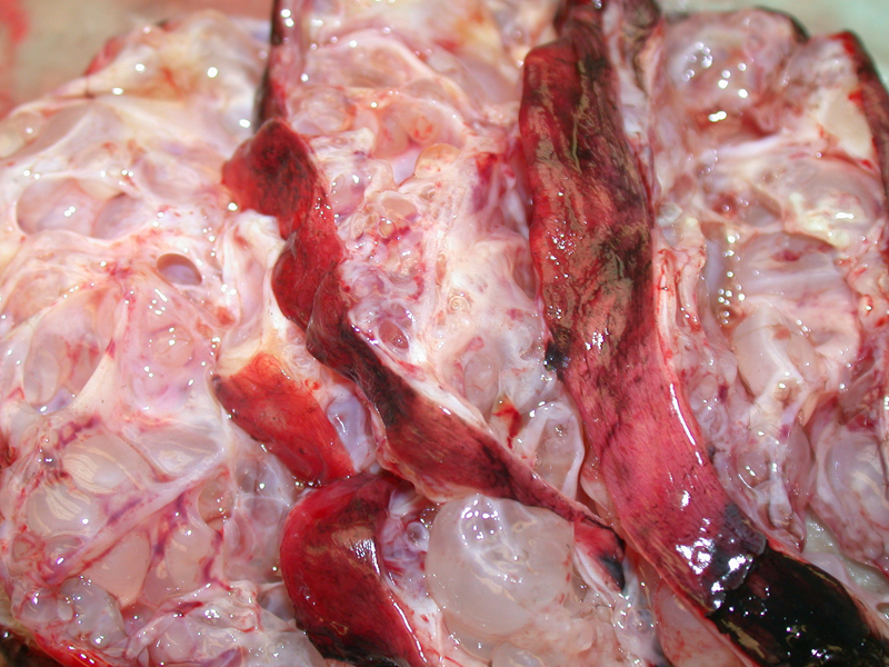
A multilocular cystic neoplasm is present. Grossly, cystic nephromas are encapsulated cystic structures located the upper pole, well demarcated from adjacent normal kidney (Cheng). Note that the cysts are simple and lack solid/papillary appearing areas. Although not shown in this image, the neoplasm is surrounded by capsule and the remainder of the renal parenchyma is normal.
)
Microscopically, the cysts are lined by flat or cuboidal epithelium which may also be hobnail as seen here. Some of the epithelium appears quite compressed. The stroma in the septa consists of dense connective tissue that may be quite cellular, resembling ovarian stroma. Interestingly, this stroma often exhibit with ER and PR receptors as demonstrated by IHC studies (Zhou).
)
Two cysts are seen along the edge of the lesion surrounded by fibrous tissue. Such lesions need to be separated diagnostically from multilocular renal cell carcinoma.
)
The edge of the lesion shows a pushing but noninfiltrative pattern of growth. The epithelial cells will stain strongly for keratins.
)
Cystic nephromas are benign cystic neoplasm usually seen in women over 30 years and male infants under 2. There are numerous cysts that are variably sized, containing a clear serous fluid.
In young children, cystic nephromas and cystic partially differentiated nephroblastoma fall under the spectrum of Wilms tumor and some consider them the same entity. These tumors have little capacity for invasion or metastasis. However, in adults, cystic nephroma do not have an association with Wilms tumor or nephrogenic rests and ought to be considered a benign biphasic tumor of stroma and epithelium. The key differential diagnosis in adults would be a multilocular clear cell renal cell carcinoma (Eble).
A bimodal distribution is seen with infant boys and middle-age women. Usually asymptomatic and found incidentally.
Benign; excision is the definitive treatment.
Cheng L, Bostwick DG, eds. Essentials of Anatomic Pathology. 2nd Ed. Totowa, NJ: Humana Press; 2006: PAGE.
Eble JN, Bonsib SM. Extensively cystic renal neoplasms: cystic nephroma, cystic partially differentiated nephroblastoma, multilocular cystic renal cell carcinoma, and cystic hamartoma of renal pelvis. Semin Diagn Pathol. 1998 Feb;15(1):2-20.
Zhou M, Magi-Galluzzi, C. Genitourinary Pathology: Foundations in Diagnostic Pathology. Philadelphia, PA: Elvesier; 2006: pAGE.