
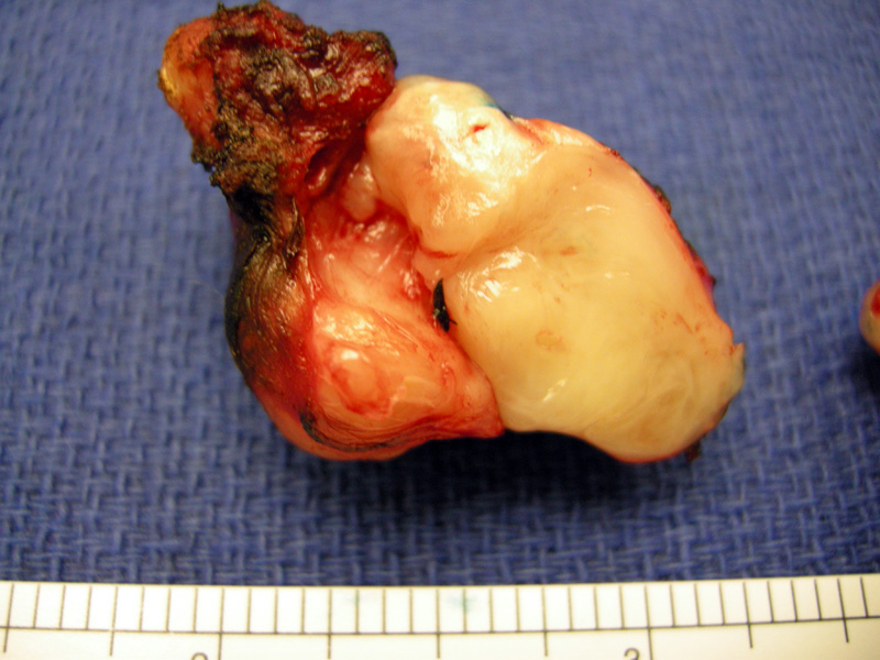
The endoscopic resection shows a mesenchymal solid mass bulging from cut surface.
)
Leiomyomas are composed of fasicles of myoid appearing bland cells with low to at most moderate cellularity. A modest amount of eosinophilic cytoplasm is seen.
)
Some areas are more hypocellular and maintain their bland appearance.
)
Foci of dystrophic calcification is also a feature.
)
Leiomyoma is seen here abutting underlying the serosa with a uniform pushing border.
)
The spindle cells stain strongly for the muscle marker desmin.
)
Strong SMA (smooth muscle actin) and lack of staining for c-kit and cd34 argue against GIST>
)
Gastrointestinal leiomyomas usually arise in the esophagus and rectum, and only rarely involve the stomach. Most gastric leiomyomas are located in the body and fundus of the stomach with a size of 1-2 cm and originate from the muscularis propria.
The vast majority have no related symptoms, and lesions are on the order of 1-2 cms. Those larger than 2 cm are more likely to have symptoms such as gastric fullness.
Benign.