
In cystitis cystica, urothelial nests acquire a cystic lumen.
)
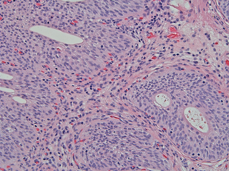
In cystitis glandularis, the cells become columnar and secrete mucin. In this nest, several glands are forming.
)
This image of cystitis glandularis demonstrates fairly complete intestinal metaplasia with goblet cell formation. In fact, the glands look very much like colonic crypts.
)
Von Brunn nests, cystitis cystica and cystitis glandularis represent a spectrum of benign morphologic changes in the urothelium. Von Brunn nests are invaginations of the urothelium, appearing as solid nests of urothelium. Some of these nests may lose continuity with the surface urothelium and appear as isolated islands of urothelium. When Von Brunn nests develop a cystic lumen, they are termed cystitis cystica. When the urothelial lining of cystitis cystica undergo glandular metaplasia, the term cystitis glandularis is used.
Many authorities believe that these changes occur as a response to inflammation and injury (similar to diverticuli in the colon). However, the fact that they are very common findings even without evidence of urothelial injury have lead some pathologists to conclude that these are simply histologic variants of normal urothelium. The association between cystitis glandularis (intestinal metaplasia) and the subsequent development of adenocarcinoma remains controversial.
1 Sternberg SS, ed. Diagnostic Surgical Pathology.4th Ed. Philadelphia, PA: Lippincott Williams & Wilkins; 2004: 1829-1835.
2 Rosai, J. Rosai and Ackerman's Surgical Pathology. 9th Ed. Philadelphia, PA: Elsevier; 2004: 1322-3.