
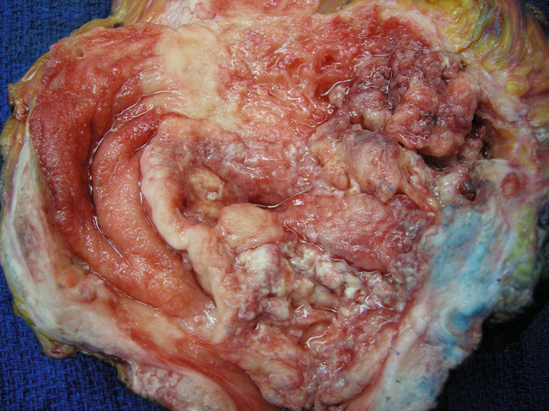
This high grade invasive TCC is bulky and friable with marked erythema. Extensive muscularis propria involvement was present.
)
High grade papillary carcinoma consists of papillary fronds lined by pleomorphic urothelial cells. There is a loss of polarity, increased nuclear size and shape variation and prominence of nucleoli. Note the discohesiveness of the cells.
)
Fusion of papillae can create solid sheets of tumor. Invasion can occur in broad sheets (as in this case), nests, clusters or single cells. In this image, confluence of cellsnalong with an inflammatory infiltrate creates a highly cellular picture.
)
Invasion into the thick muscular bundles indicative of the muscularis propria is clearly evident.
)
Highly pleomorphic cells dissect into the muscle fibers of the muscularis propria (detrusor muscle).
)
Urine cytology of high grade TCC shows enlarged cohesive cells with hyperchromatic chromatin and irregular nuclear contours.
)
A mixture of inflammatory cells and neoplastic cells is demonstrated here. Note the clumped chromatin pattern of the tumor cell
)
Pleomorphic nuclei (differing size and shape) can be appreciated in this clump of tumor cells.
)
UroVysion FISH assay demonstrates the loss of 9p21 (labeled in gold). Note that the left nucleus contains two gold signals, but the right nucleus contains none. Invasive urothelial carcinoma exhibits numerous genetic aberrations including losses in 9p21 (location of the P16 tumor suppressor gene) and duplication of chromosomes 3, 7 and 17. UroVysion FISH has been demonstrated by several studies to be more sensitive than urine cytology for detection of all stages and grades of bladder cancers. It is now used as a diagnostic test, as well as a way to monitor progression of disease.3 Note, however, FISH analysis may not be helpful in distinguishing between transitional cell carcinoma and the less common squamous cell carcinoma or adenocarcinomas of the bladder.
)
This image demonstrates aneuploidy. Pericentromeric regions of chromosome 3 are labeled in red, chromosome 7 labeled green and chromosome 17 in aqua. Basically, there are too many copies of chromosomes 3, 7 and 17. There should only be two copies per nucleus.
)
Yet another FISH image to drive home the point. Some cells lack the gold labeled 9p21, and some cells contain too many copies of chromosome 3, 7 and 17.
)
High-grade papillary urothelial carcinoma is a neoplasm of the urothelium characterized by neoplastic obviously atypial urothelial cells lining papillary fronds. Note that not all high-grade papillary carcinomas are invasive, but a significant proportion of them (15 to 40%) are. Hence, pathologists generally add the qualifier invasive or non-invasive high-grade papillary carcinoma to clearly communicate the status of invasion.
Invasion beyond the basement membrane is most important when the tumor reaches the muscularis propria. Once the muscularis propria is breached, the 5 year mortality rate lowers to 50%. Note that invasive transitional cell carcinomas can arise from flat lesions (carcinoma in-situ) or papillary lesions.
These tumors have a male predilection (approximately three times more common in men) and classically presents with painless hematuria. Cigarette smoking, exposure to aniline dyes (an aromatic amines, chemical used in manufacture of certain plastics) and chronic abuse of analgesics containing phenacetin (a compound with structural similarities to cocaine) are important risk factors.2
For stage 1 disease, transurethral resection with or without adjuvant chemotherapy is recommended. In stage 2-4, total cystectomy and prostatectomy may be necessary.
In some analyses, less than 5-10% of low-grade papillary carcinomas invade, but up to 80% of high-grade papillary carcinomas invade. Again, the actual numbers differ in depending on the study, so, just know that the majority of high-grade papillary carcinomas are invasive. Aggressive lesions can extend beyond the bladder wall and involve adjacent structures such as the prostate, ureters and retroperitoneum. Lymph node involvement and hematogenous spread to liver, lung and bone marrow occurs in advanced disease.1
• Bladder : Transitional Cell Carcinoma, Low Grade
• Bladder : Transitional Cell Carcinoma, High Grade Invasive (Case 2)
• Bladder : Urothelial Carcinoma in Situ
• Bladder : Transitional Cell Carcinoma, Low Grade
• Bladder : Transitional Cell Carcinoma, Nested Variant
• Bladder : Transitional Cell Carcinoma, Glandular Differentiation
1 Kumar V, Abbas AK, Fausto N. Robbins and Cotran Pathologic Basis of Disease. 7th Ed. Philadelphia, PA: Elsevier; 2005: 1028-33.
2 Zhou M, Magi-Galluzzi, C. Genitourinary Pathology: Foundations in Diagnostic Pathology. Philadelphia, PA: Elvesier; 2006: 172-8.
3 Halling KC, Kipp BR. Bladder cancer detection using FISH (UroVysion assay). Adv Anat Pathol 2008 Sept;15(5):279-86.