
Cells regarded as Image
)
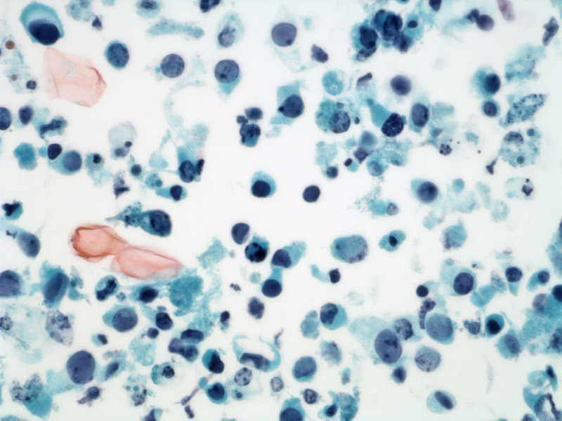
There is also cytoplasmic granularity. Many cells are seen in the urine due to loss of intercellular cohesion.
)
Viral infection is usually due to BK virus (95%) and occasionally to JC virus infection (5%) of the renal allograft. Approximately 50-80% of humans are seropositive although clinical diseases in immunocompetent hosts is extremely rare. Overall, however, approximately 5-8% of patients with renal allografts will develop interstitial nephritis.
Infection often results in asymptomatic acute or chronic graft dysfunction which may arise at any time, usually after at least 6 weeks post-transplant, up to many years later. Typically infection is within 6-12 months after transplantation.
Currently no specific treatment, although reduced immunosuppression may allow for clearance the virus.
Graft failure rates can be as high as 50–80% depending on the degree of inflammation. Biopsies with less renal scarring at the time of diagnosis are more likely to undergo resolution of the infection, in response to decrease of immunosuppression (Drachenberg).
Drachenberg CB, et al Histological patterns of polyomavirus nephropathy: correlation with graft outcome and viral load. Am J Transplant. 2004 Dec;4(12):2082-92.