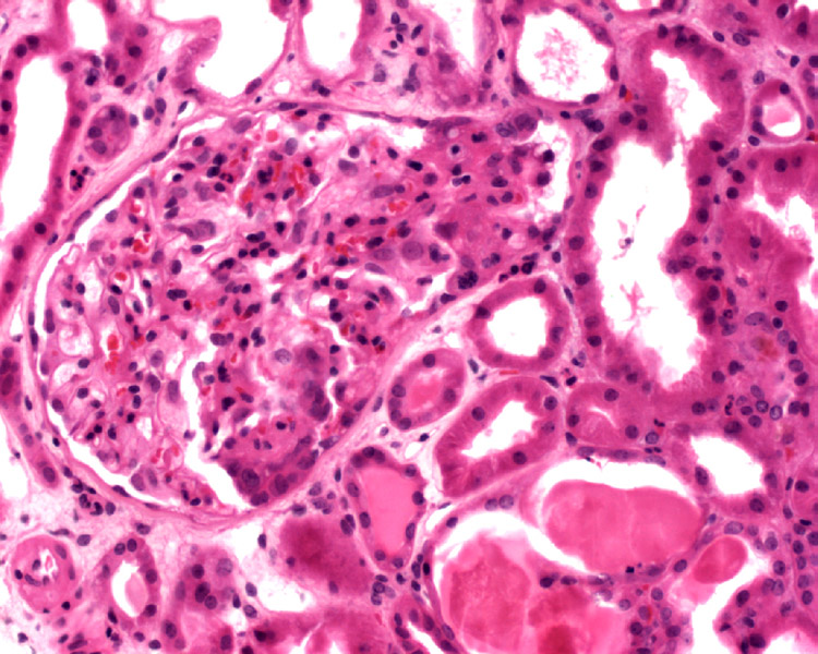

This image shows an area of consoliation at the tubular pole of the glomerulus. Although not obvious here, vacuolated foam cells may be seen along with protein resorption droplets in the lesion.
An arrow points to the origin of the proximal tubule, which appears continuous with the consolidated and sclerotic tuft.
Glomerular tip lesion is a variant of focal segmental glomerulosclerosis where the consolidated glomeruluar tuft lies adjacent to the origin of the tubular pole or synechia formation between the glomerular tuft and Bowman's capsule at the tubular pole.
Basically, the sclerotic glomerular tuft is located opposite of its usual location in classic FSGS. Note that in the glomerular tip lesion of FSGS, all the lesions must be at this tip location. If a biopsy demonstrates both tip and perihilar lesions, it is not considered a glomerular tip lesion.
It is controversial whether glomerular tip lesion represents a distinct entity or simply a common response to heavy proteinuria (Howie; Haas). In patients with glomerular tip lesion at initial biopsy, classic focal segmental glomerulosclerosis subsequently has been frequently diagnosed on repeated renal biopsy; it has also been associated with membranous and IgA nephropathy.
The clinical significance of the tip lesion remains controversial. In one retrospective review, glomerular tip lesion was identified as a distinctive and prognostically favorable clinicopathologic entity for which presenting features and outcome more closely approximate those of minimal change disease (Stokes).
Another study found a less favorable clinical outcome for this lesion. In this study by Huppes et al, they found that among 5 patients with proteinuria between 5 and 23 g/d with biopsy that showed only glomerular tip lesions, only 1 patient showed decreased proteinuria with corticosteroid and/or cyclosporine A therapy, none had complete remission of proteinuria, and 2 progressed to end-stage renal disease (Huppes). This study concluded that nephrotic syndrome characterized histologically by glomerular tip lesions is clinically indistinguishable from FSGS, and the glomerular tip lesion often should be included among histological variants of FSGS.
• Kidney : Focal Segmental Glomerulosclerosis, Collapsing Glomerulopathy
• Kidney : Focal Segmental Glomerulosclerosis
Howie A.J., Brewer D.B., The glomerular tip lesion: A previously undescribed type of segmental glomerular abnormality. J Pathol (1984) 142 : pp 205-220.
Haas M., Yousefzadeh N., Glomerular tip lesion in minimal change nephropathy: A study of autopsies before 1950. Am J Kidney Dis (2002) 39 : pp 1168-1175.
Huppes W, Hene RJ, Kooiker CJ: The glomerular tip lesion: A distinct entity or not? J Pathol 154:187-190, 1988
Kumar V, Abbas AK, Fausto N. Robbins and Cotran Pathologic Basis of Disease. 7th Ed. Philadelphia, PA: Elsevier; 2005: 962-4.
Rosai, J. Rosai and Ackerman's Surgical Pathology. 9th Ed. Philadelphia, PA: Elsevier; 2004: 1171-3.
Stokes M.B., Markowitz G.S., Lin J. et al, Glomerular tip lesion: A distinct entity within the minimal change disease/focal segmental glomerulosclerosis spectrum. Kidney Int (2004) 65 : pp 1690-1702.
Zhou M, Magi-Galluzzi, C. Genitourinary Pathology: Foundations in Diagnostic Pathology. Philadelphia, PA: Elvesier; 2006: 354-8.