System: Head and Neck: Ear: Neoplastic: Inverted Schneiderian Papilloma

System: Head and Neck: Ear: Neoplastic: Inverted Schneiderian Papilloma

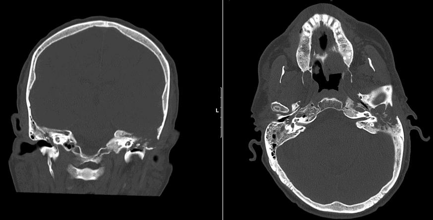
These CT scans demonstrate an osteolytic process with erosion of the tegmen tympani and mastoid bone.
de Filippis and colleagues (2002) reported a case of inverted papilloma involving the middle ear and mastoid, which prompted a literature search of this rare entity. They found that only 12 cases of inverted papillomas of the middle ear (temporal bone) have been reported thus far.
Inverted papilloma can arise from extension of a pre-existing sinonasal papilloma or as a primary tumor, arising from ectopic Schneiderian mucosa. Interestingly, there are some major epidemiologic and etiologic differences between inverted papillomas of the middle ear and ones arising from sinonasal passages. From their examination of 13 cases, de Filippis found that there is a female predominance (vs. male predominance in sinonasal inverted papillomas) and HPV infection was found in only 12% of cases (vs. up to 76% in sinonasal inverted papillomas). Furthermore, inverted papillomas of the middle ear appear to be more aggressive with higher risk of malignant transformation, usually into squamous cell carcinoma (de Filippis).
Presenting symptoms include unilateral conductive hearing loss, pain, or discharge. In a review of 5 patients with middle ear inverted papillomas, three patients had intact tympanic membranes. The remaining two patients had perforated tympanic membranes through which a polypoid mass could be seen (Wenig).
• Sinonasal : Schneiderian Papilloma, Oncocytic Type
• Sinonasal : Schneiderian Papilloma, Inverted Type
de Filippis C, Marioni G, Tregnaghi A, Marino F, Gaio E, Staffieri A. Otol Neurotol. Primary inverted papilloma of the middle ear and mastoid. 2002 Jul;23(4):555-9.
Wenig BM. Schneiderian-type mucosal papillomas of the middle ear and mastoid. Ann Otol Rhinol Laryngol. 1996 Mar;105(3):226-33.
)