
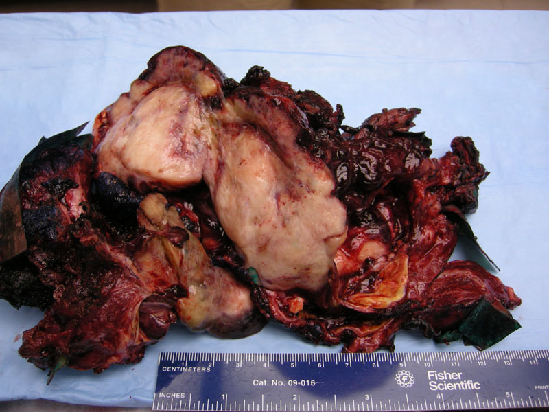
Tumor excision is the mainstay of treatment. In this surgical resection, bulky fleshy solid tan nodules of tumor are seen.
)
A primitive appearing neoplasm is seen with ovoid cells set in a pale staining background with intervening vessels.
)
The tumor is characterized by malignant mesenchymal stroma which is shown here, and is usually accompanied by blastematous and sarcomatous elements.
)
Cystic components are lined by modified respiratory epithelium, and the surrounding stroma is myxoid and modestly cellular.
)
This image represents the cystic type, which clinically and by imaging may mimic CCAM.
)
A different example of this tumor shows frankly malignant cells with a blunt spindled cell shape.
)
This tumor was associated with marked fibrosis, a feature which may be variably present.
)
This third example shows more pronounced cellularity surrounding vessels.
)
Tumors are subclassified as type I (purely cystic), type II (cystic and solid), or type III (purely solid). MRI may demonstrate solid enhancing nodules inside fluid-filled cavities, a mass causing lung compression, mediastinal shift, and pleural effusion. No preoperative imaging can reliably differentiate between congenital cystic lesions and PPB type I.
About 25% of patients are found to have another lesion such as pulmonary cysts, cystic nephromas, sarcomas, medulloblastomas, thyroid dysplasias and neoplasias, malignant germ cell tumors, Hodgkin disease, leukemia, and Langerhans cell histiocytosis.
These tumors usually develop in young children without congenital pulmonary malformations. They may present with symptoms attributable to an upper respiratory infection, or possibly with a pneumothorax. The mean age for type I at presentation = 10 months, with type II and III arising in older children.
Therapy involves total resection of the tumor, even in those with microscopic residual disease. The role of combination chemotherapy has not been defined on a statistical basis, but it is commonly regarded that chemotherapy should be considered. In some studies, adjuvant chemotherapy appears to benefit type I PPB patients (Priest 2006).
Prognosis is poor because of local recurrence and metastases (Parsons). The 2-year survival is 83% for type I, 49% for type II and 42% for type III, even with multimodality treatment (surgery, chemotherapy and radiotherapy). However, the overall survival rate is 45% at 5 years, with no significant difference between histological types (Priest 1997)
Priest JR et al. Type I pleuropulmonary blastoma: a report from the International Pleuropulmonary Blastoma Registry. J Clin Oncol. 2006 Sep 20;24(27):4492-8.
Parsons SK, Fishman SJ, Hoorntje LE et al (2001) Aggressive multimodal treatment of pleuropulmonary blastoma. Ann Thorac Surg 72:939–942
Priest JR, McDermott MB, Bathia S et al (1997) Pleuropulmonary blastoma: a clinicopathologic study of 50 cases. Cancer 80:147–161