System: Serosal Membranes: Peritoneum: Malignant: Mesothelioma Overview

System: Serosal Membranes: Peritoneum: Malignant: Mesothelioma Overview

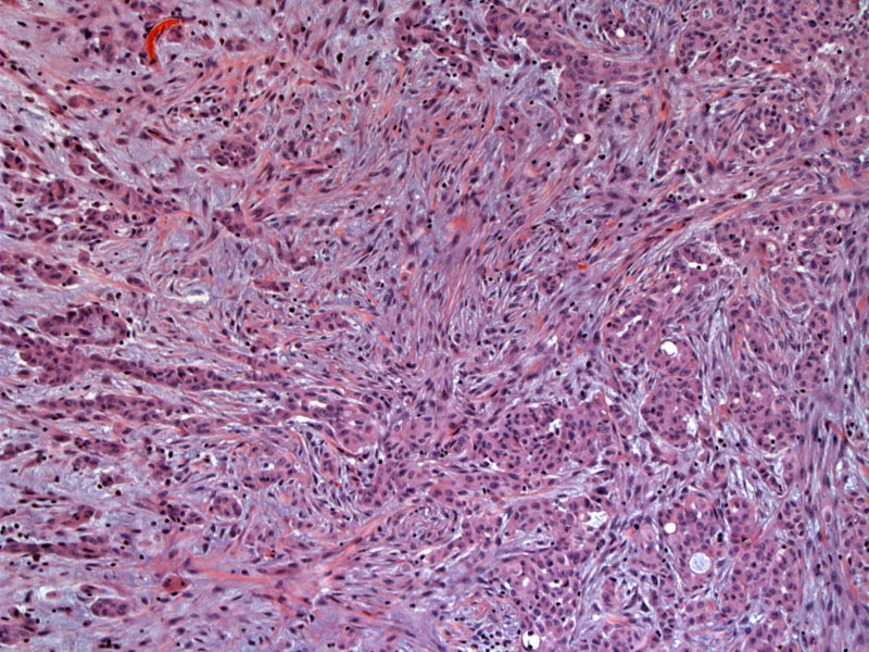
Irregular epithelioid cells in small groups lie in a fibromyxoid backgroud that contains small vessels. Image
The cells are mesothlial and are haphazardly arranged in a cohesive manner. Some glandular appearing lumina are apparent. Image
Small clusters, some of which appear gland like, are seen. In some areas the cells line up and form a glomeruloid shape. Image
Strong nuclear WT-1 staining is evident. Image
calretinin highlights the infiltrating mesothelioma Image
Mesotheliomas can assume a wide range of morphologies, and thus, a broad range of differential diagnoses must be considered (as noted in the above chart).
Epithelioid mesotheliomas are composed of polygonal or round cells with a paracentric nuclei, a small nucleoli and a moderate amount of cytoplasm.
Butnor KJ. My approach to the diagnosis of mesothelial lesions. J Clin Pathol. 2006 Jun;59(6):564-74.
Cagle PT, Churg A. Differential diagnosis of benign and malignant mesothelial proliferations on pleural biopsies. Arch Pathol Lab Med. 2005 Nov;129(11):1421-7.
Churg A, Colby TV, Cagle P et al. The separation of benign and malignant mesothelial proliferations. Am J Surg Pathol. 2000 Sep;24(9):1183-200.
)
)
)
)
)