
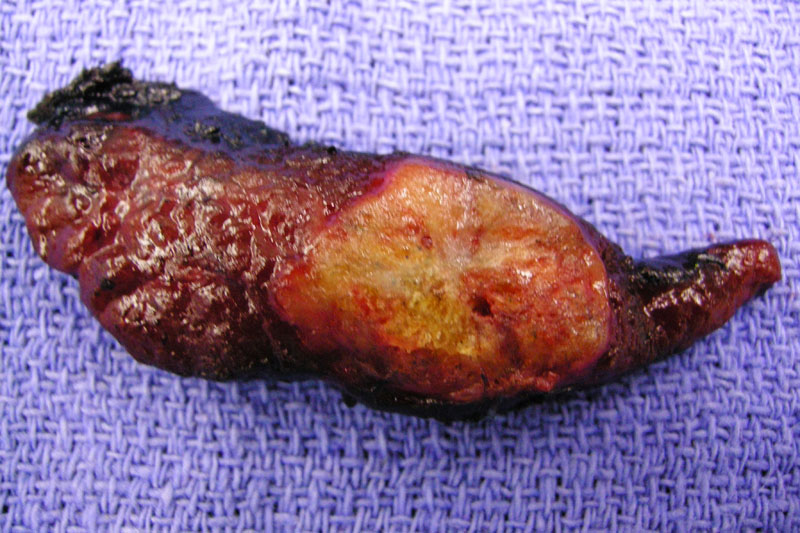
The lobe contains a fairly well circumscribed yellow-red medullary carcinoma with areas of calcification.
Medullary thyroid carcinoma can grow in a variety of patterns (solid sheets, nested/organoid, trabecular, papillary etc...) In this image, the neoplastic C-cells appear to be percolating through, forming thin trabeculae. Several vascular lumenina are seen, but no normal follicles are present.
This is a more organoid pattern of growth.
The tumor cells here have a more spindled morphology. The typical salt and pepper chromatin pattern can be appreciated.
Cells are engulfed by large geographic zones of amyloid deposits. Congo red and birefringence confirmed these deposits as amyloid.
The neoplastic parafollicular C cells demonstrate strong staining for calcitonin.
Being neuroendocrine cells, they will also exhibit positivity with chromogranin or synaptophysin.
Note also, that the neoplastic parafollicular cells will be TTF-1 positive, but thyroglobulin negative (as compared to papillary thyroid carcinomas in which both TTF-1 and thyroglobulin would be positive).
Aspiration cytology of medullary carcinoma illustrates loose clusters of spindle cells with indistinct cytoplasmic borders.
Pap stain highlights the amorphous amyloid material.
The cell block shows ovoid to spindled cells around a vascular network. The nuclear features of a neuroendocrine differentation are clearly evident. The cytoplasm almost takes on an oncocytic look.
This different patient underwent an FNA which yield a cellular neoplasm composed of short spindle cells. Many single cells are present.
The nuclear chromatin is uniform and nucleoli are not prominent.
The cells on the cell block stain nicely for calcitonin.
Medullary thyroid carcinoma (MTC) is a neuroendocrine neoplasm composed of parafollicular C-cells, which elaborate calcitonin. 75% of these tumors are sporadic and 25% are associated with an inherited syndrome. Sporadic tumors tend to be unilateral and solitary, whereas familial tumors tend to be bilateral, multicentric and present at an earlier age.
Familial MTCs are associated with MEN syndromes, which are autosomal dominant syndromes with endocrine manifestations. MTCs is most strongly associated with MEN Type 2A (Sipple Syndrome), defined as MTC, pheochromocytomas and parathyroid hyperplasia. MEN Type 2B consists of MTCs, pheochromocytomas, mucosal neuromas and a Marfanoid body habitus.
Patients with familial syndromes have germline mutations of RET (located on chromosome 10q11.2) whereas 50% of sporadic cases have a somatic mutation of RET. The specific mutation of RET leads to a different phenotype. For example, MEN2B patients with the M918T mutation have a more aggressive form of MTC compared to MEN2A and FMTC, which share a variety of mutations in other codons (Moo-Young).
Parafollicular C-cell hyperplasia can be physiologic or pre-neoplastic lesion to the familial forms of MTC. Criteria to distinguish between these two forms is not clear. Using the definition in the Lester text, greating than 50 parafollicular C cells constitute a pre-neoplastic lesion (Thompson). In the Fletcher text, aggregates of parafollicular C cells should be solid, replacing normal thyroid follicles with mild atypia. Either way, know that C-cell hyperplasia can exist as a response to normal aging, renal disorders or thyroiditis, OR it can be a precursor lesion to the hereditary type of MTC (with germline RET proto-oncogene mutations).
Grossly, these tumors tend to be nonencapsulated and firm. They are usually located at the upper half of the gland since this is where the C cells reside. Histologically, polygonal cells form trabeculae and are surrounded by hyalinized stroma and amyloid (which may be composed in part of calcitonin). Other architecture patterns are seen as well and include glandular, pseudopapillary and paraganglioma (zellballen)-like patterns. MTC is immunoreactive for calcitonin, chromogranin, synaptophysin CEA, TTF-1, among others.
MTC accounts for about 5-10% of all thyroid cancers and is clinically significant because a high percentage of those patients have a germline mutation of RET and thus, may have MEN2A, MEN2B or FMTC. Genetic testing should be offered to these patients.
Presents with a thyroid mass, compressive symptoms such as dysphagia, hoarseness and cervical lymphadenopathy. Patients may experience diarrhea and flushing if they have very high levels of calcitonin and other peptide hormones (ie. serotonin) secreted by this tumor.
Surgery is the mainstay as these tumors are not sensitive to chemotherapy or radiation.
The overall 5, 10 and 15-year survival rates of 65-87%, 51-78% and 65%, respectively (Thompson). Clinical stage is an important prognostic factor. Note that over ~50% present with cervical lymph node metastasis.
Survival appears to be best with the familial MTC (a hereditary form not associated with other endocrinopathies), followed by MEN 2A and sporadic MTC. MEN 2B have the poorest prognosis (with the highest percentage of patients presenting with distant metastasis)(Thompson, Fletcher).
→MTC arises from parafollicular C cells of the thyroid.
→The familial syndromes to consider are MEN2A, MEN2B and non-MEN familial medullary carcinoma (FMTC).
→The responsible gene in familial cases of MTC is the germline mutation of RET. There are somatic mutations of RET in about 50% of sporadic cases.
• Thyroid : C Cell Hyperplasia
• Thyroid : Hyalinizing Trabecular Adenoma
Fletcher CDM, ed. Diagnostic Histopathology of Tumors. 3rd Ed. Philadelphia, PA: Elsevier; 2007:1038-1045.
Moo-Young TA, Traugott AL, Moley JF. Sporadic and familial medullary thyroid carcinoma: state of the art. Surg Clin North Am. 2009 Oct;89(5):1193-204.
Thompson LDR. Endocrine Pathology: Foundations in Diagnostic Pathology. Philadelphia, PA: Elsevier; 2006:113-122.
Late in the course of the disease, the tumor tends to metastasis to lung, liver, adrenals and bone. For more information regarding this entity, please view our case for metastatic medullary thyroid carcinoma to the liver

)
)
)
)
)
)
)
)
)
)
)
)
)
)