
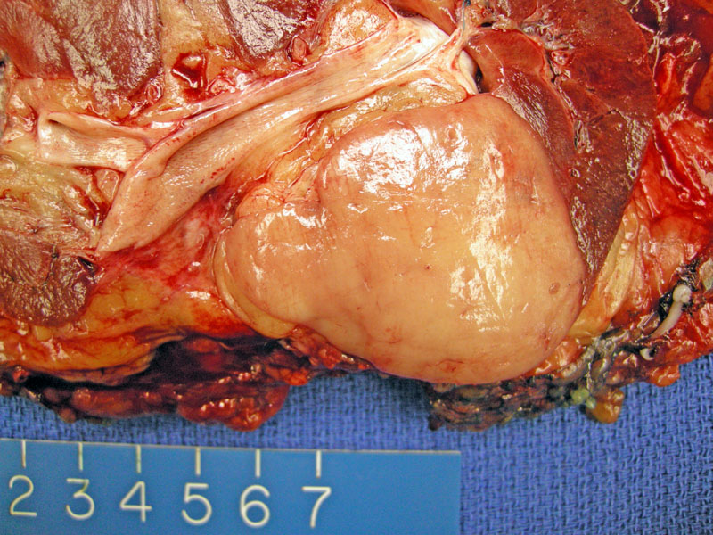
Metanephric adenoma is well circumscribed but not encapsulated, and uniform throughout.
)
Metanephric adenomas are composed of tightly-packed small round blue cells arranged in compact tubules. Note the sharp distinct border with the adjacent kidney (on the right).
)
The neoplastic cells are arranged in nests. The stroma is usually inconspicuous. Although not clearly demonstrate here, a papillary structures may protrude into a cystic lumen, creating glomeruloid structures.
)
Another image demonstrates that small round blue tumor cells are arranged in nests. The scant stroma can be edematous or hyalinized.
)
The neoplasm is composed of primitive, small, round, blue cells with delicate chromatin and inconspicious nucleoli. These cells are just slightly larger than lymphocytes. Mitoses should not be present.
)
Metanephric neoplasms consist of three groups: metanephric adenomas, metanephric stromal tumors, and metanephric adenofibromas. Metanephric adenomas are composed of epithelial cells; metanephric stromal tumors are composed of spindled fibroblastic cells; metaphric adenofibromas are a mixed of epithelial and stromal elements.
Historically, the two key entities to be distinguished from metanephric adenomas were Wilm's tumors and papillary renal cell carcinomas (PRCC). The small round cells in compact tubular structures in metanephric adenomas were morphologically similar to the epithelial (and sometimes blastema) component of Wilms tumors, and the papillary structures variably present in metanephric adenomas were morphologically similar to type 1 PRCCs. Note that type 1 PRCCs consist of small, basophilic cells and type 2 PRCCS consist of larger, polygonal cells with abundant eosinophilic cytoplasm.
Recent studies have indicated that metanephric adenomas bear histogenetic similarities to Wilms tumors and may in fact be the mature, differentiated form of Wilms tumor. An analogous sequence of tumors would be neuroblastomas (immature), ganglioneuroblastomas and ganglioneuromas (mature, benign and inactive). Thus, the sequence for Wilms spectrum of tumors may be nephrogenic rests (immature), Wilms tumors and metanephric neoplasms (mature, benign and inactive).2
The close relationship of metanephric adenomas and Wilms tumors is supported by the fact that metanephric adenomas stain positively for WT1, which encodes a tumor suppressor and transcription activator essential for the proper development of genitourinary structures. Although WT1 positivity has been demonstrated in non-renal tumors (e.g. desmoplastic small round cell tumor, mesothelioma), WT1 positivity in renal tumors is limited to nephroblastic tumors (Wilms tumors), nephrogenic rests, cystic partially differentiated nephroblastoma, rhabdoid tumor and now, metanephric adenomas.3
Metanephric adenomas are distinguished from type 1 papillary renal cell carcinomas (PRCC) by key chromosomal differences. PRCC is characterized by trisomies of chromosome 7 and 17, stain strongly for CK7 and keratin AE1, and are negative for WT1 and CD57. In contrast, MAs do not have abnormalities in chromosome 7 and 17, stain strongly for WT1 and CD57 and are negative for CK7 and keratin AE1. MAs and PRCCs can also be distinguished by a number of histologic features. For example, PRCCS have characteristic papillary cores with foamy macrophages.
Although metanephric adenomas are the most common epithelial renal neoplasm in children and young adults, most of these tumors occur in adults (peak incidence 5th to 6th decade). There is a clear female predilection with a F:M ratio of 2:1. Usually asymptomatic, but 10-15% of patients present with polycythemia vera.1
Excision is curative.
Excellent as tumor is benign.
1 Zhou M, Magi-Galluzzi C. Genitourinary Pathology: Foundations in Diagnostic Pathology Philadelphia, PA: Elvesier; 2007: 304-5.
2 Argani P. Metanephric neoplasms: The Hyperdifferentiated, Benign End of the Wilms Tumor Spectrum? Clinics in Laboratory Medicine 25 (2005) 379-392.
2 Muir TE et al. Metanephric Adenoma, Nephrogenic Rests, and Wilm's Tumor: A Histologic and Immunophenotypic Comparions. Am J Surg Pathology (2001) 25(10): 1290-1296.
Please visit our Wilm's Tumor case to learn more about nephroblastic tumors.