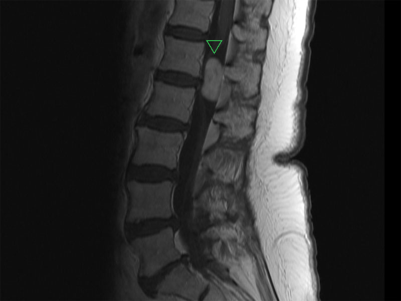

MRI shows a soft tissue mass (arrow) within the spinal canal posterior to the cauda equina at the level of L1-2. There is associated mass effect on the cauda equina. Possibilities include a glial tumor, schwannoma, or meningioma.
Cells with round to ovoid nuclei and granular chromatin are embedded in a fibrillary background. They tend to form rosettes, gland-like structures or canals. Perivascular pseudorosettes are particularly characteristic, with the fibrillary processes of the cells extending toward the lumen of a vessel.
Low power view highlights dilated blood vessels and perivascular pseudorosettes. Pseudorosettes are composed of tumor cells radially arranged around blood vessels with a zone of anuclear fibrillary processes. Pseudorosettes are not as specific for ependymoma as true rosettes and ependymal canals. True ependymal rosettes and canals contain columnar cells arranged around a central lumen (not seen here).
Classic ependymoma is composed of sheets of monomorphic cells with round to oval nuclei and poorly defined cellular borders. Nuclei have are a salt and pepper distribution, but are not coarse (as seen in astrocytomas or oligodendrogliomas). Nucleoli are inconspicuous.
Areas with regressive changes are common. In this view, numerous pigmented macrophages containing hemosiderin are present. Other types of regressive changes that can be seen are myxoid degeneration, calcifications, hemorrhage and sometimes focal areas of cartilage and bone formation. Areas of non-palisading necrosis (not seen here) are still compatible with WHO grade 2.
GFAP stains the majority of the tumor cells but not the vessels.
The touch prep shows uniform cells with ovoid nuclei and a modest amount of pink cytoplasm.
Ependymoma is a glial neoplasm that displays ependymal differentiation. Most ependymomas arise next to the ventricular system lined by ependyma, which includes the central canal of the spinal cord (Kumar). Ependymomas account for 3% to 9% of all brain tumors. 90% arise in the brain and 10% arise in the spinal cord (Prayson). In adults, spinal ependymomas are the most common spinal neuroepithelial tumor, comprising 50 to 60% of all spinal gliomas (Ellison, Fletcher).
Ependymomas in the posterior fossa are usually intraventricular (fourth ventricle) and form solid papillary masses whereas supratentorial ependymomas are periventricular and tend to be more cystic as well as anaplastic (Prayson). Cystic degeneration, calcifications, hemorrhage and necrosis may be present. Although well-demarcated with "pushing borders", complete excision is often not possible since the tumor is in close proximity to the pontine and medullary nuclei of the brainstem. Complete excision is more feasible in spinal ependymomas (Kumar).
Ependymomas are strongly and diffusely positive for GFAP (glial fibrillary acidic protein) and have variable staining for EMA (membrane and/or cytoplasmic dot-like staining) and CAM5.2 (Fletcher).
The most common cytogenetic aberration is monosomy of chromosome 22 (occuring in 1/3 of tumors). Loss of 6q and gain of 1q is especially common in anaplastic ependymomas (Fletcher).
Subtypes of ependymoma include: cellular, papillary, clear cell and tanycytic. Myxopapillary ependymoma is a distinct entity with a more indolent clinical course (WHO grade I tumor).
Ependymoma is most commonly a slowly growing neoplasm seen in children and younger adults; however, they are known to occur in all age groups. The fourth ventricle is the preferred site in the children, while in adults, the spinal cord is the favored location. Ependymomas in the spinal cord are often seen in the context of NF2.
Posterior fossa (e.g. fourth ventricle) tumors can lead to hydrocephalus due to obstruction of the fourth ventricle and CSF flow.
In our particular case, however, a man in his early 30's complained of headache, nausea and vomiting for approximately one month. MRI revealed a 4th ventricular enhancing heterogeneous obstructing mass with tumoral extension into the medial and lateral apertures. The tumor measured 4.4 cm in greatest dimension. He also complained of a 2 year history of hiccups.
Tumor excision followed by external beam radiation therapy.
Prognosis is guarded. Due to the relationship with the ventricular system, CSF dissemination is common. Survival after resection and radiotherapy is about 2 years (Kumar). Outcome for spinal cord and supratentorial ependymomas is better compared to posterior fossa tumors.
Most are well-differentiated and considered WHO Grade II. Anaplastic transformation (e.g. high mitotic activity, necrosis, dedifferentiation) can occur and are WHO Grade III tumors with more aggressive behavior.
• Brain : Ependymoma, Anaplastic Variant
• Brain : Ependymoma, Myxopapillary
Cheng L, Bostwick DG, eds. Essentials of Anatomic Pathology. 2nd Ed. Totowa, NJ: Humana Press; 2006: 375-6..
Ellison D, Love S, Chimelli L, et al.(2004). Neuropathology: A reference text of CNS pathology, 2nd ed. New York: Mosby.
Kumar V, Abbas AK, Fausto N. Robbins and Cotran Pathologic Basis of Disease. 7th Ed. Philadelphia, PA: Elsevier; 2005: 1404-5.
Louis D, Ohgaki H, Wiestler O, Cavenee W.(2007). WHO Classification of Tumors of the Central Nervous System 4th ed. Lyon: International Agency for Research on Cancer (IARC)