
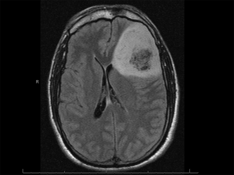
MRI shows a large mass lesion in the left frontal lobe. The mass appears intracranial and shows minimal contrast enhancement.
)
Another MRI image of the same lesion shows central necrosis and moderate mass effect most consistent with an intermediate or high grade glioma. This is a grade III anaplastic oligodendroglioma.
)
Anaplastic oligodendrogliomas (WHO grade III) have increased cellularity when compared to a grade II oligodendroglioma.
)
Increased cellularity and nuclear pleomorphism can be appreciated here as well.
)
A different case shows some compartmentalization of cells, which grow in sheets with pseudocystic spaces.
)
The cells show nuclear anaplasia and a solid growth pattern.
)
GFAP stains all the cells strongly.
)
MAP2 is similarly strong.
)
Focal WT-1 is present.
)
Ki-67 stains about 20% of the cells.
)
Here is yet another case showing significant nuclear atypia and an atypical mitotic figure.
)
Anaplastic oligodendrogliomas (WHO grade III) have the following histopathologic features: (1) increased mitotic activity defined as greater than 6 mitoses per 10 HPF; (2) hypercellularity; (3) endothelial proliferation; (4) pleomorphism; (5) an epitheliod morphology as the cells tend to have increased cytoplasm with more distinct cell borders; (6) prominent nucleoli; and (7) areas of micronecrosis. MIB-1 labeling may be helpful in identifying anaplastic tumors when elevated (Prayson, Ellison).
Prognosis for grade II oligodendrogliomas is quite good, with average survival time of 10-15 years. Those with the codeletion of 1p and 19q may survive for even longer as these tumors are extremely responsive to PCV (procarbazine, CCNU, vincristine) chemotherapy. Anaplastic oligodendrogliomas (grade III) have an average survival time of 3-5 years, although some patients with 1p/19q tumors may survive longer (Prayson).
Ellison D, Love S. Neuropathology: A Reference Text of CNS Pathology. 2nd Ed. London, UK: Mosby; 2004; 641-4.
Prayson, RA. Neuropathology: Foundations in Diagnostic Pathology. Philadelphia, PA: Elvesier; 2005: 457-465.