System: Gastrointestinal: Colon: Pre-Neoplastic: Tubular Adenoma

System: Gastrointestinal: Colon: Pre-Neoplastic: Tubular Adenoma

The polyp head shows crowded adenomatous glands characterized by enlarged elongated nuclei and a normal amount of intervening lamina propria.
While clearly adenomatous in nature, some such polyps retain and even exaggerate their cytoplasmic mucin.
Uncommonly the glands may show dilated lumens and irregular contours.
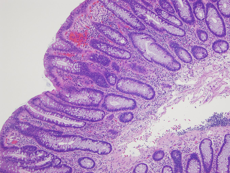
Neutrophils infiltrate the surface epithelium of this polyp. It is not uncommon for polyps to become secondarily inflamed, a feature which should not be overinterpreted as a primary inflammatory process.
Another polyp with extensive acute inflammation within the nodular protrusion of the adenoma.
Finding a lymphoid follicle in a biopsy obtained for a bump.
Occasionally a non-invasive lesion may simulate malignancy, as seen here in this large adenomatous polyp involving the cecum.
another example Image
This adenoma contains lots of Paneth cells, which can be seen in those arising from the right colon.
Nice uniform pseudostratified nuclei are seen, lining crypts of various shapes.
• Colon : Adenocarcinoma, Conventional Type
• Colon : Tubulovillous Adenoma
• Colon : Adenoma with Pseudoinvasion
)
)
)
)
)
)
)
)
)
)