
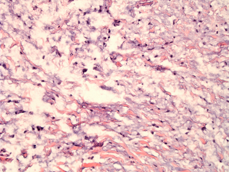
The typical features of a myxoma are present in this lesion, with a loosely arranged stellate or spindle-shaped cells proliferation within a myxoid matrix.
)
Small stellate cells are present within a bit of delicate stroma. Cellularity is rather low and atypia is absent. Tumor cells express vimentin and muscle specific actin, if one were to investigate.
)
Odontogenic myxoma (OM) is defined as a benign odontogenic tumor of mesenchymal origin that shows locally invasive behavior and consists of rounded and angular cells that lie in abundant mucoid stroma.
It is a slow-growing tumor with the potential to attain considerable size without noticeable signs and symptoms. In fact, such tumors can grow to involve half of the maxilla or mandible including the ramus and the condyle.
The tumor causes teeth displacement. Radiologically, there are multilocular lesions with trabeculations, described as "tennis racket,” “soap bubble,” or “honeycomb” to depict the internal structure. In one study, the mean age was 31 years (Noffke).
Conservative therapy results in a high recurrence rate, so en bloc resection may be indicated.
This is considered to be a benign odontogenic tumor but with locally aggressive behavior.
Leiser Y, Abu-El-Naaj I, Peled M. J. Odontogenic myxoma--a case series and review of the surgical management.Craniomaxillofac Surg. 2009 Jun;37(4):206-9.
Noffke CE, Raubenheimer EJ, Chabikuli NJ, Bouckaert MM. Odontogenic myxoma: review of the literature and report of 30 cases from South Africa. Oral Surg Oral Med Oral Pathol Oral Radiol Endod. 2007 Jul;104(1):101-9.