
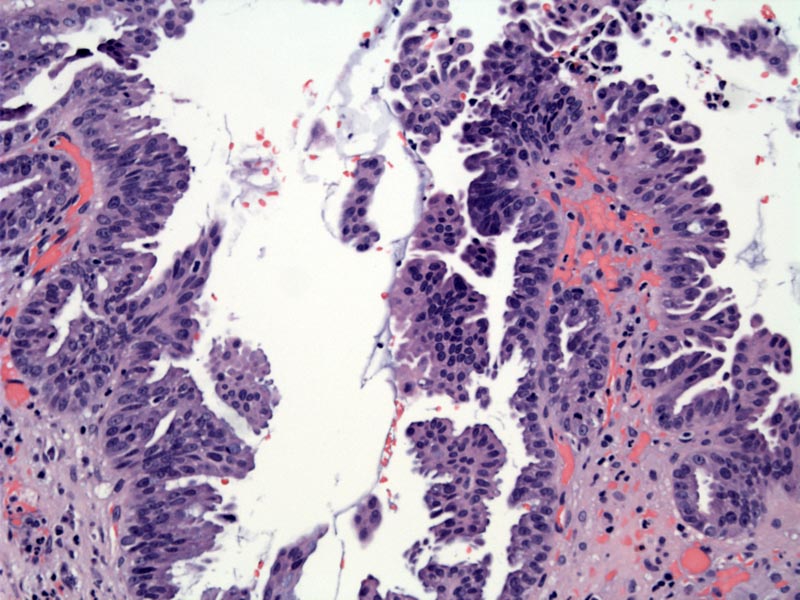
The surface epithelium is replaced by a villous architecture harbouring high grade epithelial cells. High grade dysplasia is demonstrated by pleomorphism, loss of nuclear polarity, and prominent nucleoli.
The malignant glands have abundant apical clear cytoplasm and variable shapes and sizes resembling those seen in pancreatic adenocarcinoma.
Other areas of the same tumor show higher grade nuclei, mucin, and irregular desmoplasia. Variability of grade within a given tumor is not uncommon.
There is often marked desmoplasia with a variable density of malignant glands.
This different case manifests tubuloglandular structures within a dense desmoplastic background.
Variability in glandular morphology is typical. One malignant gland is large while others are small.
A different case shows compact trabecular architecture.
Peritoneal washing shows a rare structure compatible a glandular structure. Note the small lymphocyte in the upper left for size comparison
The cells are most often present in the washing as single cells, which could be easily overlooked.
The cell block shows single cells with eccentric nuclei.
Gallbladder carcinoma is the 4th most common malignancy in the GI tract. Native Americans and Hispanics are disproportionately represented. Over 80% of patients have gallstones (Fletcher). Chronic irritation and epithelial damage to the mucosa leads to proliferation and increased risks for mutation, and thus are risk factors for neoplastic change. Other associated risks for gallbladder carcinoma include porcelain gallbladders, anomalous pancreaticbiliary ductal junction (APBDJ) and familial adenomatous polyposis.
Porcelain gallbladder (diffuse dystrophic calcifcations) is strongly associated with gallbladder carcinoma. Although porcelain gallbladder is rare (0.5% of cholecystectomies), 20% of them contain gallbladder carcinoma (Iacobuzio). Anomalous pancreaticbiliary ductal junction (APBDJ), more frequently seen in Japan, occurs when the pancreatic and common bile duct join outside of the duodenal wall where the sphincter of Oddi is located. Thus, there is an increased risk of reflux of pancreatic secretions up the biliary tract (Fletcher, Iacobuzio).
Grossly, the gallbladder wall is usually diffusely thickened. Rarely, one might see an intraluminal papillary growth. Sometimes, a gallbladder carcinoma presents as subtle thickenings of the wall that can easily be mistaken for chronic cholecystitis on examination. Most adenocarcinomas arise from flat dysplasia and not from pre-existing adenomas (Iacobuzio). Currently, the sequence of carcinogenesis is thought to be metasplasia (especially intestinal) to dysplasia to carcinoma (Iacobuzio).
Histologically, invasive glands are lined by columnar or cuboidal cells resembling gallbladder epithelium. Goblet cells are commonly found. The degree of atypia often is market, and exceeds what one what expect for the degree of gland formation. The neoplastic glands can be arranged in sheets, cords, glands or in cribriform patterns. The amount of desmoplasia is variable and an inflammatory infiltrate is often seen. These tumors usually demonstrate positivity with CK 7, CEA and keratins. CK20 positivity is variable.
Variants include: clear cell (must distinguish from metastatic RCC), adenosquamous (dual glandular and squamous differentiation), undifferentiated (spindled and giant cells similar to anaplastic pancreatic carcinoma), signet ring (mimics lobular carcinoma of breast), small cell neuroendocrine (oat cell) and papillary (tumor cells proliferate on a fibrovascular stalk and connotes a better prognosis).
Affects older adults (peak in the 7th decade) with a female predilection (F:M ratio of 2:1). Symptoms may be similar to cholecystitis in early disease; in advanced disease, there may be anorexia, weight loss and jaundice. Unfortunately, patients tend to present at an advanced stage.
Most undergo routine cholecystectomy because the diagnosis is unsuspected. Hepatic resection and lymphadenectomy after diagnosis may still benefit those with relatively low stage tumors. In retrospective analysis, findings from >2,000 patients with adenocarcinoma of the gallbladder who had undergone surgery demonstrated that those who received adjuvant radiotherapy had superior overall survival, but the survival advantage appeared to be restricted to patients with node-positive disease and tumors invading the liver (Mojica).
Poor prognosis, similar to their pancreatic counterparts. Overall survival is on the order of 10%, with pathological stage being the most important prognostic indicator.
• Gallbladder : Clear Cell Adenocarcinoma
Fletcher CDM, ed. Diagnostic Histopathology of Tumors. 3rd Ed. Philadelphia, PA: Elsevier; 2007: 445-6.
Iacobuzio-Donahue CA, Montgomery EA. Gastrointestinal and Liver Pathology: Foundations in Diagnostic Pathology. Philadelphia, PA: Elsevier; 2005: 441-451.
Mojica P, Smith D and Ellenhorn J. Adjuvant radiation therapy is associated with improved survival for gallbladder carcinoma with regional metastatic disease, J Surg Oncol 96 (2007), pp. 8–13.

', 'micro', ''))
)
)
)
)
)
)
)
)
)