
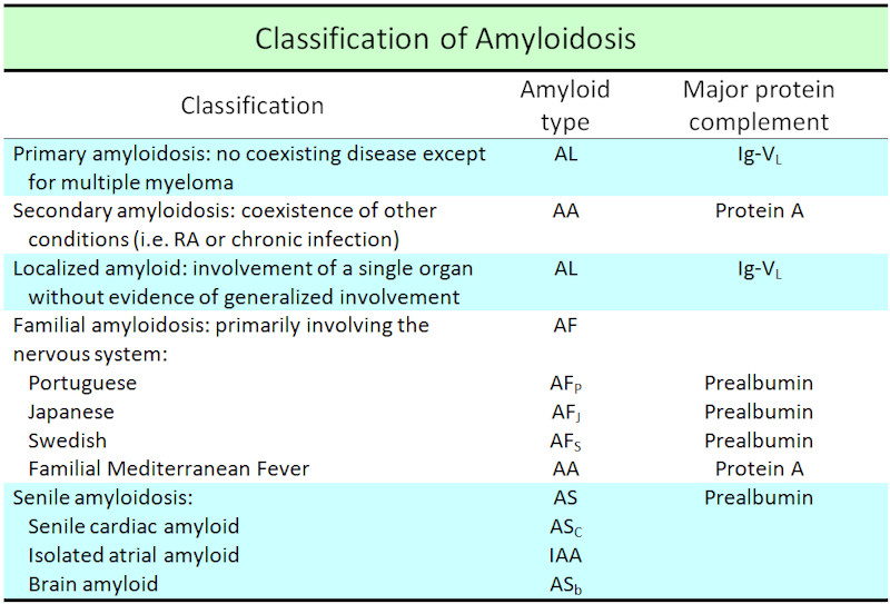
Classification scheme for amyloidosis adapted from reference 2.
)
The more eosinophilic area are skeletal muscle fibers, with separation due to the infiltration of amyloid, which is within the expanded intermuscular area.
)
Deposition of amyloid between muscle fibers is prominent, resulting in macroglossia. Loss of the submucosal glands is often seen as well.
)
Congo Red highlights confluent areas of amyloid deposition as bright red. There is perivascular amyloid staining as well.
)
Polarization does show apple green birefringence but is not easily photographed.
)
Amyloid disease is an umbrella term for a group of conditions characterized by interstitial deposition of a proteinaceous fibrillar material, amyloid. In reality amyloid is composed of fibrils containing low molecular weight protein subunits of plasma Over 20 different human subunit proteins have been identified.
Several different subtypes of amyloidosis occur, some hereditary (autosomal dominant) mutations in protein precursors.
Types of Amyloidosis
- AL amyloidosis: Due to protein deposition from Ig light chain fragments produced from plasma cells. AL amyloid is sometimes found in association with multiple myeloma.
- AA amyloidosis: Due to protein deposition fibrils composed of fragments of the acute phase reactant serum amyloid A. This is typically seen in cases of chroinic disease with recurring inflammation such as RA or chronic infections.
- Dialysis related amyloidosis: Due to deposition of fibrils derived from beta-2 microglobulin.
- Senile amyloidosis: Due to deposition of otherwise normal transthyretin in myocardium and other sites
- Organ/Tissue specific amyloidosis: Due to deposition of precursor protein which are synthesized and processed at sites contiguous to amyloid deposition. Alzheimer's disease is an example of this with deposition of beta protein (Ab).
As detailed above, the deposit of amyloid can be localized or systemic. The localized form presents minor clinical manifestation (e.g. hoarseness or dysphagia) when compared to the large organ involvement seen in systemic disease, and local amyloid may not be progressive. Localized amyloidosis affecting the head and neck region is an uncommon and benign process. The most common sites of involvement are the larynx, subglottis, and thyroid [3]. Unlike laryngeal amyloidosis, involvement of the tongue usually signals primary (systemic) amyloidosis. As such, involvement of the tongue should prompt evaluation for possible systemic disease.
In cases of organ specific amyloid deposition, such as in this case, massive involvement can significantly impair function. Reduction surgery may be needed, however, with function severely compromised. Persistent or recurrent disease is a problem, however.
Depends of the severity of organ involvement. The median survival of those with primary (systemic) amyloidosis is on the order of 1-2 years.
1 Pentenero M, Davico Bonino L, Tomasini C, Conrotto D, Gandolfo S. Localized oral amyloidosis of the palate. Amyloid. 2006 Mar;13(1):42-6.
2 Nandapalan V, Jones TM, Morar P, Clark AH, Jones AS. Localized amyloidosis of the parotid gland: a case report and review of the localized amyloidosis of the head and neck. Head
Neck 1998;20:73–78.
3 UpToDate online 17.2 accessed 8/24/09. "An overview of amyloidosis". http://www.uptodate.com/online/content/topic.do?topicKey=othrheum/13441&selectedTitle=1~150&source=search_result