System: Bone: Vascular: Benign: Aneurysmal Bone Cyst


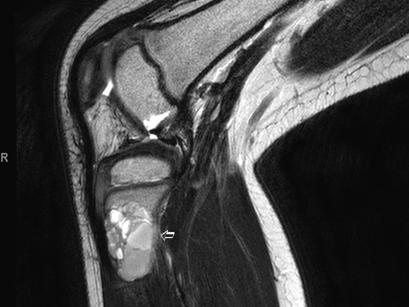
MRI shows a heterogeneous lesion in the proximal tibia. Need more description here. Image
cystic spaces filled wtih RBCs, solid patternless stroma with mononucleated and osteoclast like giant cells Image
2nd case, fibular lesion Image
cystic spaces Image
another area Image
osteoclast like giant cells in more solid foci Image
)
)
)
)
)
)