System: CNS: Spinal cord: Neoplastic: Intramedullary schwannoma

System: CNS: Spinal cord: Neoplastic: Intramedullary schwannoma

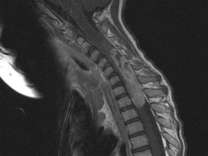
MRI shows a large mass centered in the spinal cord extending predominantly posteriorly.
spindled fascicular Image
thick walled vessels; some veracoy bodies to right Image
pink cyto, some nuclear variability, resembles meningioma a bit but is EMA negative Image
diffuse positivity for S100 Image
)
)
)
)
)