System: Soft Tissue: Myxoid: Neoplastic: Low Grade Fibromyxoid Sarcoma

System: Soft Tissue: Myxoid: Neoplastic: Low Grade Fibromyxoid Sarcoma

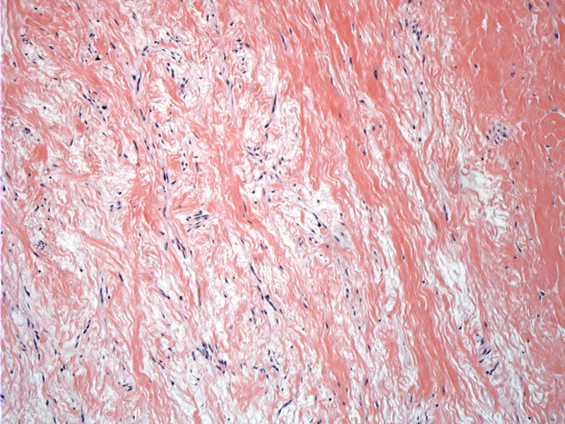
The tumor consists of myxoid stroma admixed with bland spindle cells irregularly infiltrating the adjacent collagenous tissue (to the right).
It is easy to tell how the tumor gets its name. Again, there are some collagen streaks present amongst a background of very bland spindle cells embedded in a bluish tinged myxoid stroma.
Intersecting fascicles of fibroblastic spindle cells in a variably fibromyxoid stroma can raise a broad differential. IHC and molecular studies would be helpful.
If you could imagine a more hyalinzed core, this may pass as a giant rosette. These structures are featured prominently in the hyalinzing spindle cell tumor with giant rosettes, a variant of low-grade fibromyxoid sarcoma.
CT scan of the tumor shows a large slightly heterogeneous mass demonstrating internal calcification and lobulation. It extends along the left visceral space displacing the left thyroid lobe anteriorly and medially and extends as far down as the mediastinum. There is a clear demarcation between the lesion and the vessels.
gross Image
new case Image
super bland Image
looks like LG fibromyxoid sarc Image
here is the whorling again in this tumor Image
ddx includes low grade myxofibrosarcoma Image
gross comparison Image
chart comparison Image
Low-grade fibromyxoid sarcoma (LGFMS, Evans tumor) should not be confused with low-grade myxofibrosarcoma (MFS), despite their near identical names. LGFMS is considered a variant of fibrosarcoma, whereas MFS is considered a separate distinct entity. Furthermore, LGFMS affects younger adults (30s to 50s) whereas myxofibrosarcomas arise in older adults (60s to 80s).
Nearly all LGFMS exhibit the distinct cytogenetic aberration t(7;16)(q34;p11) This translocation results in the fusion gene FUS-CREB3L2 (Panagopoulos, Fletcher). Myxofibrosarcoma does not have a defining chromosomal translocation and has complex cytogenetic aberrations.
In terms of histology, myxofibrosarcoma has curvilinear vessels and spindle/oval cells in a myxoid background, with variable numbers of pseudolipoblasts. LGFMS is composed of spindle cells, often arranged in a whorled pattern. There may be a distinctive alternating pattern to the myxoid and fibrous stroma. Compared to myxofibrosarcoma, the fibrous and collagenous component is much more prominent and the myxoid component less so. The curvilinear vessels typical of MFS is not seen in LGFMS. Furthermore, in MFS, higher grade lesions exhibit increasing cytologic atypia, whereas atypia is minimal in LGFMS.
While the neoplastic spindle cells in LGFMS react for vimentin, they are usually negative for antibodies to keratin, desmin, muscle specific actin, S100 protein, CD34, CD31, or epithelial membrane antigen. This IHC profile is helpful in distinguishing LGFMS from myofibroblastic or schwannian lesions (Fletcher).
A variant, termed "hyalinizing spindle cell tumor with giant rosettes" (Lane), consists of bland spindle cells, with fibromyxoid areas but also contains hyalinized acellular islands surrounded by oval and spindle cells in a palisading pattern. This variant exhibits the same translocation t(7;16)(q34;p11) as low-grade fibromyxoid sarcoma.
A wide age range may be affected but they are most common in young adults. These tumors most often occur in the lower limb or groin region but occasionally are encountered in other deep soft tissues.
Despite its bland appearance, LGFMS does have metastatic potential. Various case series have reported different statistics in terms of prognosis. For example, the original report by Evans found that 7 out of 12 cases developed distant metastatis (Oda). However, other studies have revealed a less aggressive course. In 54 cases where follow-up (2-192 months) was available, 5 patients had recurrences, 3 metastases, and 1 death (Folpe).
→LGFMS affecsts younger adults (30s to 50s) whereas myxofibrosarcomas arise in older adults (60s to 80s).
→A distinct cytogenetic aberration t(7;16)(q34;p11) has been identified in these tumors.
→Hyalinizing spindle cell tumor with giant rosettes is a variant that exhibits the same t(7;16) translocation.
• Uncertain Lineage : Sclerosing Epithelioid Fibrosarcoma
• Myxoid : Myxofibrosarcoma, Low Grade
Fletcher CDM, ed. Diagnostic Histopathology of Tumors. 3rd Ed. Philadelphia, PA: Elsevier; 2007: 1556.
Folpe AL, Lane KL, Paull G, Weiss SW. Low-grade fibromyxoid sarcoma and hyalinizing spindle cell tumor with giant rosettes: a clinicopathologic study of 73 cases supporting their identity and assessing the impact of high-grade areas. Am J Surg Pathol. 2000 Oct;24(10):1353-60.
Lane, K. L. , R. J. Shannon , and S. W. Weiss . Hyalinizing spindle cell tumor with giant rosettes: a distinctive tumor closely resembling low-grade fibromyxoid sarcoma. Am J Surg Pathol 1997. 21:1481–1488.
Oda Y, et al. Low-grade fibromyxoid sarcoma versus low-grade myxofibrosarcoma in the extremities and trunk. A comparison of clinicopathological and immunohistochemical features. Histopathology 2004, 45, 29-38.
Panagopoulos, I. , C. T. Storlazzi , and C. Fletcher . et al. The chimeric FUS/CREB3L2 gene is specific for low-grade fibromyxoid sarcoma. Genes Chromosomes Cancer 2004. 40:218–228.
Vernon SE, Bejarano PA. Low-grade fibromyxoid sarcoma: a brief review. Arch Pathol Lab Med. 2006 Sep;130(9):1358-60.

)
)
)
)
)
)
)
)
)
)
)
)
)