System: Soft Tissue: Chondroid: Benign: Soft Tissue Chondroma

System: Soft Tissue: Chondroid: Benign: Soft Tissue Chondroma

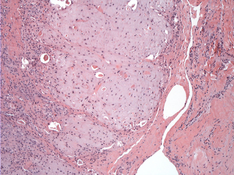
The lesion is to the left, with lobulated hypocellular hyaline cartilae, and the well demarcated margin to the right, with normal soft tissue to the far right. This was a 1 cm lesion. Image
Part of the lesion is calcified. In additional to seconary calcification, some lesions may become myxoid or fibrous. Image
some variable cellularity Image
)
)
)