System: Head and Neck: Salivary Gland: Malignant: Low Grade Cystadenocarcinoma

System: Head and Neck: Salivary Gland: Malignant: Low Grade Cystadenocarcinoma

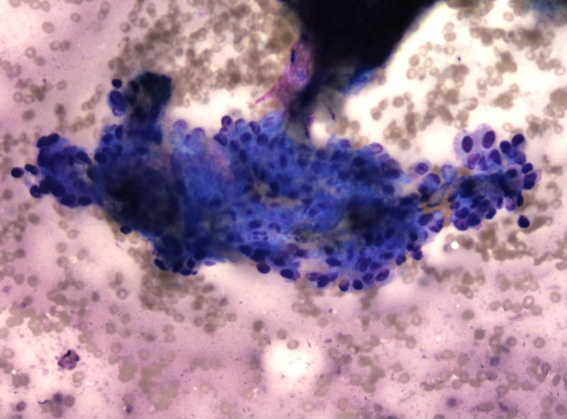
Ovoid basaloid cells in a sheet comprise the FNA, and are devoid of metachromatic mesenchyma fragments.
The irrregular cluster of basaloid cells lack atypia and mitoses, and are present within a bloody background.
Ribbon like growth in a hyalinized stroma are widespread, without areas of mesenchymal differentiation.
Large areas showed this pattern, which suggests basal cell adenoma.
another area Image
The tumor grows within a cystic structure lined by these same cells
The cyst wall appears simple, with irregular areas of solid growth within the cavity
An unencapsulated area is appreciated with adjacent normal parotid tissue.
Closer inspection confirms tumor cells mingling with fat, indicative of a low grade malignancy.
• Salivary Gland : Low grade Mucoepidermoid Carcinoma
)
)
)
)
)
)
)
)
)