System: Head and Neck: Oral Cavity: Inflammatory: Drug Eruption

System: Head and Neck: Oral Cavity: Inflammatory: Drug Eruption

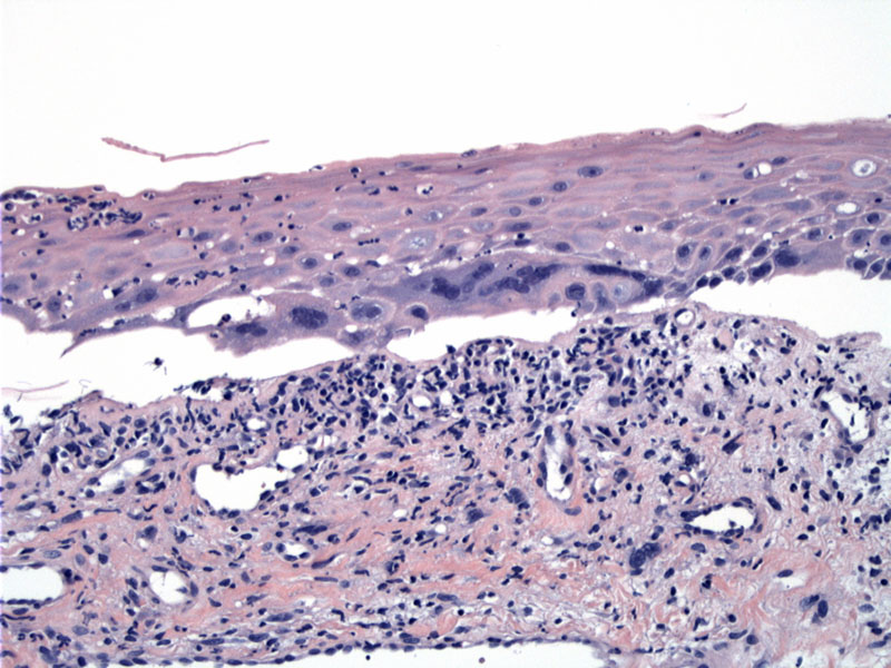
This is a biopsy of the left cheek which shows separation of the epithelium and marked basal hyperchromasia and multinucleation. Image
Atypical basal zone cells are seen and there is a reactive capillary proliferation resembling granulation tissue in the stroma. Neutrophils infiltrate the epithelium. Image
Focal ulceration is seen. Image
)
)
)