System: Bone: Chondroid: Neoplastic: Osteochondroma

System: Bone: Chondroid: Neoplastic: Osteochondroma

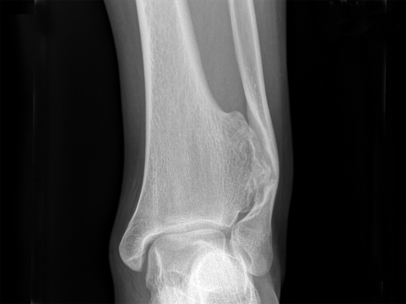
Plain films show a sessile osteochondroma projection off of the distal tibial metaphysis. It is deforming the distal fibular metadiaphyseal region.
The lesion recapitulates the growth plate, seen here as a broad-based protuberance composed of a cartilage cap (pale bule) that undergoes endochondral ossification to make trabecular bone (deep pink).
The cartilage layer containing chondrocytes in their lacunae is lined by a fibrous cap.
A cartilaginous cap contains chondrocytes, which form orderly columns toward the base of the lesion which transitions into a zone of endochondral ossification. Fat and marrow elements may be admixed with the ossified areas.
Osteochondroma, a common benign cartilage-forming tumor of bone, arises in the metaphysis of long bones in adolescents or young adults. Most arise as a solitary and sporadic lesion. It is thought that this is imply aberrant growth of bone, where the bone grows away from the growth line instead of in line with it.
Multiple lesions may occur in patient with hereditary multiple exostoses in which there is a germline mutation of EXT1 or EXT2 (tumor suppressor genes).
On imaging, osteochondroma is seen as an outward protrusion from native bone. The neoplasm is contiguous with underlying bone and can be sessile or pedunculated (with a stalk) i.e. the cortex and spongiosa of the tumor is continuous with the cortex and spongiosa of the underlying non-neoplastic bone.
Peak incidence in the second or third decade and with a male predominance (M:F of 2:1). It usually presents as a painless bump near the joints. The most common sites are the distal femoral, proximal tibial or proximal humeral metaphyses.
Small tumors are asymptomatic, but larger lesions may cause pain by infringing on adjacent structures or due to fracture of the stalk (Folpe).
Rapid growth of a preexisting osteochondroma suggest malignant transformation, which occurs in about 1%-3% of tumors. Those with multiple osteochondroma are at a much higher risk of malignant transformation (5%-25%)(Folpe).
→Most are sporadic. Multiple osteochondromas can arise in the setting of hereditary multiple exostoses, in which there are germline mutations of EXT1 or EXT2.
→The tumor is exophytic and covered by a cartilage cap.
→Histologically recapitulates the epiphyseal growth plate.
Fletcher CDM, ed. Diagnostic Histopathology of Tumors. 3rd Ed. Philadelphia, PA: Elsevier; 2007: 1602-3.
Folpe AL, Inwards CY. Bone and Soft Tissue Pathology: Foundations in Diagnostic Pathology Philadelphia, PA: Elsevier; 2010: 330-2.
)
)
)
)