
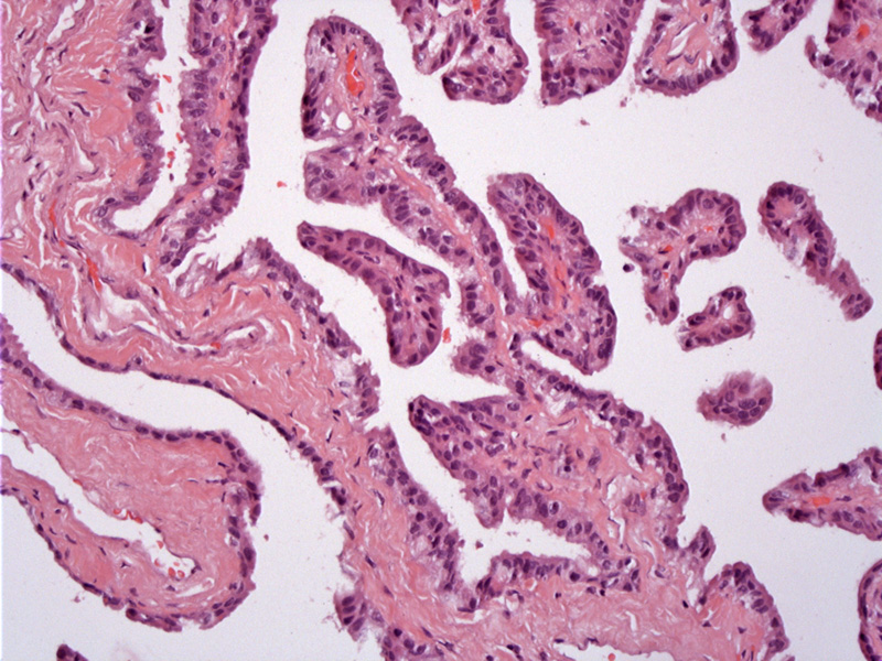
Well-developed papillomatous proliferations are lined by a relatively bland single layer of cuboidal cells. The vascularized papillae shows some stromal collagenization.
)
Psammomatous bodies are common findings. There should be no significant mitotic activity or pleomorphism.
)
The choroid plexus is a specialized structure composed of modified ependymal cells that produce CSF, which then flows through the ventricular system, bathing the brain and spinal cord.
Most neoplasms of the choroid plexus are choroid plexus papillomas (CPP), however, a few will become atypical (atypical CPP) or malignant (choroid plexus carcinoma).
Grossly, CPPs are well-circumscribed with a stippled papillary surface. Histologically, CPPs resemble normal choroid plexus, but with more exuberant growth. Papillary projections with fibrovascular cores are lined by columnar epithelium. Common to both CPPs and CPCs are dystrophic psammomatous calcifications and xanthoma cells (Ellison). Occasionally, the lining cells may exhibit cilia.
In atypical CPPs and CPCs, there is increasing cytolgic atypia and architectural complexity. Mitotic activity is also increased as highlighted by Ki-67 labeling. In CPC, there is invasion of the stroma and necrosis.
Most CPPs arise in children (over 50% present before age 2) and are located in the lateral ventricles. The minority that arise in adults tend to be in the fourth ventricle. Due to blocked CSF drainage as well as increased CSF production, hydrocephalus is the most common symptom.
CT findings are those of a well-defined lobulated mass with an irregular frond-like pattern, resulting in a cauliflower-like appearance.
Surgical excision.
Ellison D, Love S. Neuropathology: A Reference Text of CNS Pathology, 2nd Ed.Philadelphia, PA: Elvesier; 2005: 685-8.