System: Gastrointestinal: Liver: Developmental: Congenital Hepatic Fibrosis

System: Gastrointestinal: Liver: Developmental: Congenital Hepatic Fibrosis

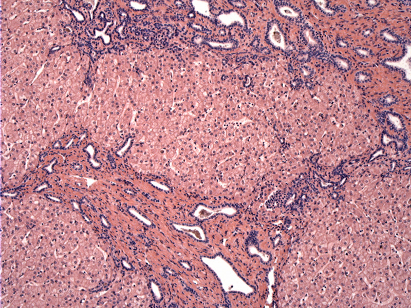
portal portal bridging fibrosis; portal tract expanded by fibrosis Image
proliferation of duct prolifles along the periphery of the limiting plate Image
bile plugs; little to no inflammation; hepatic parenchyma shows no giant cell change or hepatocyte alterations; hepatic cholestasis is absent Image
staghorn shaped duct prolifes; buboidal epithelium; absent reactive ductular proliferation, rather these are primary malformed ducts Image
Nice gross image Image
interlacing pattern Image
Arrest of embyrologic remodeling of the ductal plate, leading to abnormally increased biliary channels forming a ring along the portal tract periphery. A centrally located bile duct is generally absent.
May be associated with polycystic kidney disease autosomoal recessive.
Clinical presentation varies but may include cytopenias, bleeding varices or splenomegaly. The liver is enlarged. The process may be patchy, making needle biopsy sometimes unreliable for diagnosis. Complications include portal hypertension, cholangitis, and cholangiocarcinoma.
)
)
)
)
)
)