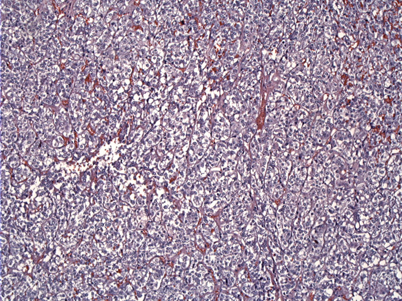

The tumor is composed of epithelioid (round/polygonal) cells arranged in sheets. The vesicular nuclei are randomly located within the cell and contain multiple prominent nucleoli. The cytoplasm is granular and slighly eosinophilic. The cell borders are quite distinct. There may be intercellular stroma composed of collagen fibers, occasionally creating an organoid pattern (Yamamoto).
Immunohistochemically, these tumors stain for smooth muscle markers (e.g. SMA, alpha-sarcomeric actin) and are negative for desmin. In general, epithelioid LMS in the soft tissues do not express desmin to the degree that conventional LMS do (Yamamoto, Suster). Ultrastructual studies confirm their smooth muscle nature (e.g. cytoplasmic thin filaments, plasmalemmal densities, etc...). This image shows the expected diffuse and strong staining for smooth muscle actin.
There is focal desmin positivity.
This second example shows epithelioid cells hwith ave distinct cell outlines. The nuclei are randomly located, although most appear to be eccentric in this case.
Epithelioid leiomyosarcoma is a variant of leiomyosarcoma characterized by round and polygonal smooth muscle cells. This tumor is usually seen in the uterus, however, there have been a handful of case reports and case series describing this entity in the soft tissues. It is important simply to be aware that epithelioid LMS exists as its appearance can mimic malignant melanoma, epithelioid sarcoma and metastatic carcinoma.
• Myometrium : Leiomyosarcoma, Epithelioid Variant
Suster S. Epithelioid leiomyosarcoma of the skin and subcutaneous tissue. Clinicopathologic, immunohistochemical, and ultrastructural study of five cases. Am J Surg Pathol. 1994 Mar;18(3):232-40.
Yamamoto T, et al. Epithelioid Leiomyosarcoma of the External Deep Soft Tissue. Arch Pathol Lab Med. 2002;126:126:468-70.