System: Skin: Adnexal: Sweat Gland Neoplasm: Syringocystadenoma Papilliferum

System: Skin: Adnexal: Sweat Gland Neoplasm: Syringocystadenoma Papilliferum

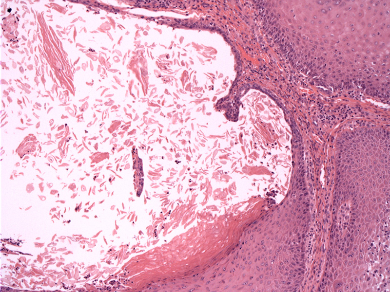
Case 1: An invaginated dilated structure contains numerous papillae, although only one lonely papilla is seen here. The papillae are lined by a two-cell epithelial layer which may exhibit decapitation (apocrine) secretion.
SCAP is considered a benign hamartoma, derived from eccrine or apocrine glands. Mutations in tumor suppressor gene p16 may play a role in its pathogenesis (Obaidat).
Histologically, it is characterized by a cystic space (connected to the epidermis) which contains papillary projections. These papillae are lined by two layers of epithelial cells (luminal columnar cells that exhibit decapitation secretions and outer cuboidal cells). Another characteristic feature is the presence of a robust plasma cell infiltrate in the stroma (Rapini, Obaidat).
SCAP commonly occurs on the scalp or face of young adults. 40% of cases are associated with a congenital lesion, usually nevus sebaceous. The typical clinical presentation is a lesion with absent hair growth is seen on the scalp. The surface may be red, granular and weepy (Fletcher).
→Cystic space within the tumor connects to the epidermis. Papillae project into the cystic space, lined by epithelial and apocrine cells. A plasma cell infiltrate is seen within the stroma.
→Commonly on seen on the scalp of young adults.
→Nevus sebaceus may be a precursor lesion.
• Vulva : Hidradenoma Papilliferum
Busam KJ. Dermatopathology: Foundations in Diagnostic Pathology 1st Ed. Philadelphia, PA: Elsevier; 2010: 403-4.
Fletcher CDM, ed. Diagnostic Histopathology of Tumors. 3rd Ed. Philadelphia, PA: Elsevier; 2007: 1454.
Obaidat NA. Skin adnexal neoplasms - part 2: An approach to tumors of cutaneous sweat glands. J Clin Pathol 2007;60:145-159.
Rapini RP. Practical Dermatopathology. Philadelphia, PA: Elsevier; 2005: 295-6.

)