
Case 1, image 1: This 47 year-old woman died of a seizure disorder. At autopsy, there was marked neuronal loss and gliosis of hippocampi and right temporal lobe.
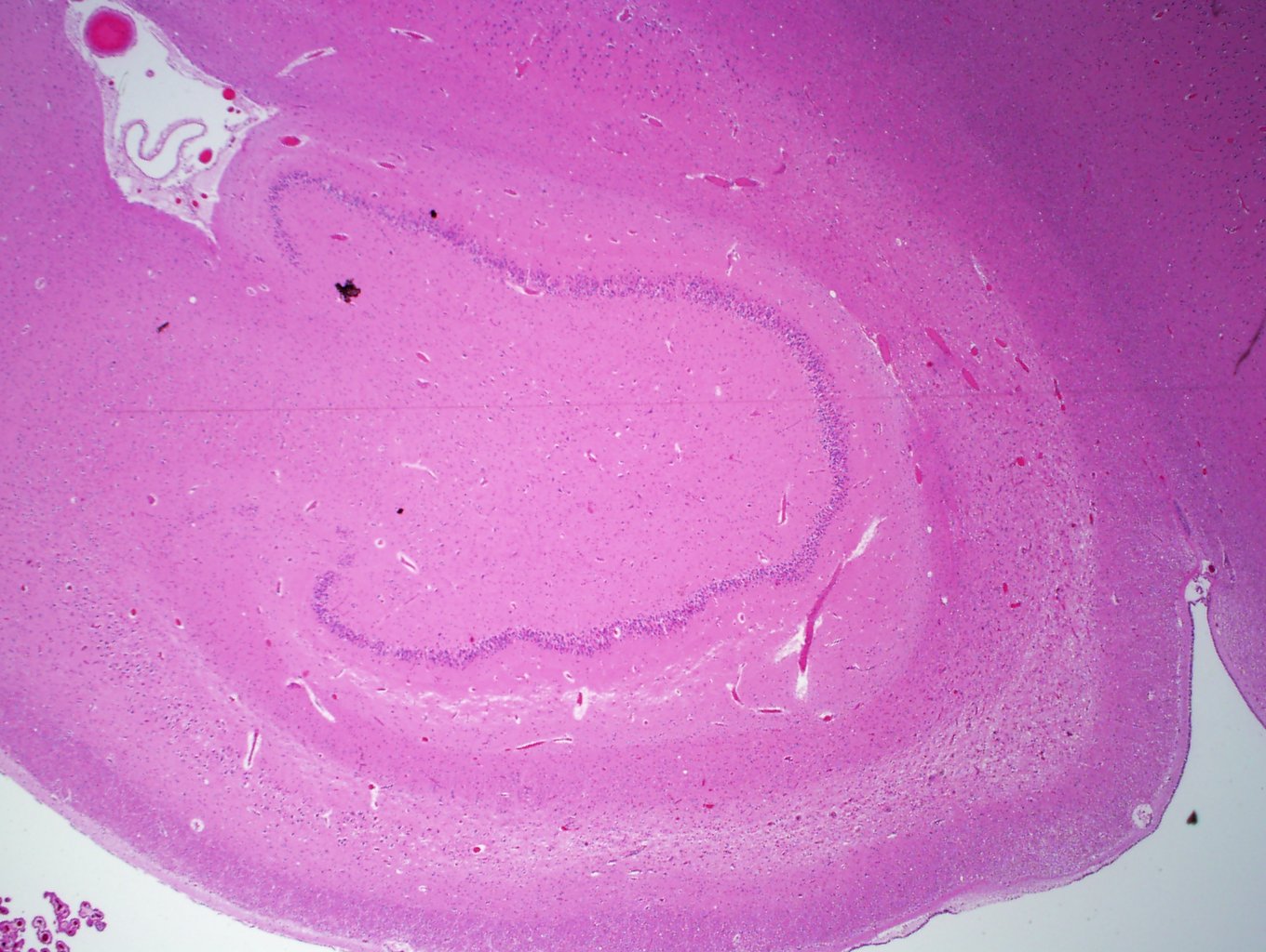
Case 1, image 2: Sections from the right hippocampus reveal marked neuronal loss and gliosis, greatest in sectors CAI, CAII and CAIII.
Case 1, image 3: Note the pallor and loss of neurons.
Case 1, image 4: Gliosis and neuronal loss. There are neurons on the right side of the image, and then, rather sudden dropout starting at the center of the image.
Case 1, image 4: Yet another image.
Hippocampal sclerosis consists of loss of neurons in the dentate nucleus and the CA1 and CA4 sectors of the hippocampus with variable gliosis. Grossly, this may be seen as shrinkage of the hippocampus.
Hippocampal sclerosis may be seen in individuals with a history of seizures in addition to individuals with Alzheimer's disease and other dementias.
Agamanolis, A. The Pathology of Seizures. NeuropathologyWeb.Org. Available at : http://neuropathology-web.org/chapter2/chapter2dseizures.html

)
)
)
)
)