
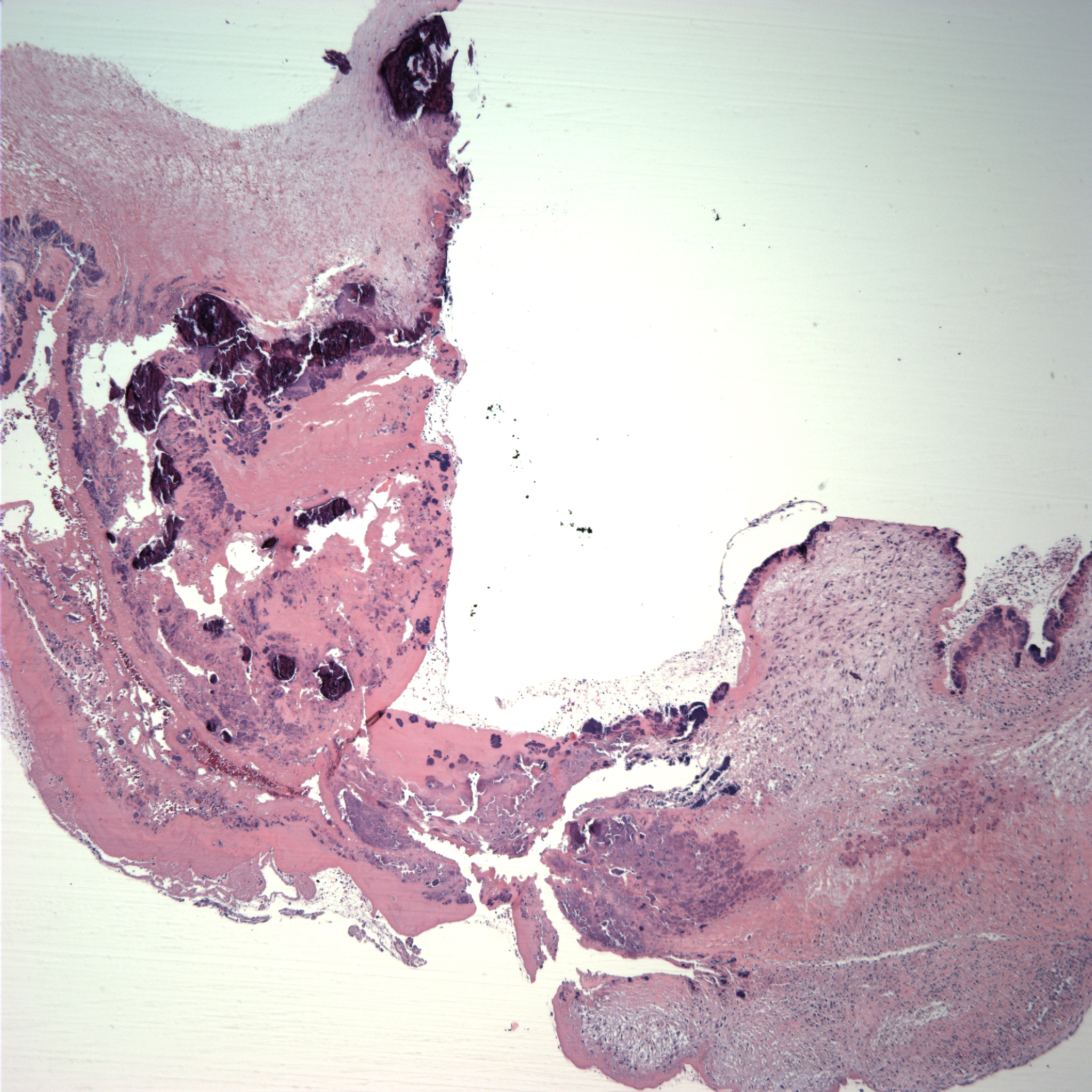
Case 1, image 1: This 31 year old man died of a bicuspid aortic valve. The autopsy revealed a congenital abnormality of the aortic valve. Specifically, the aortic valve had two, instead of three, cusps. This resulted in obstruction of blood outflow from the heart. His heart consequently was markedly enlarged and he had evidence of heart failure. A section of the valve demonstrates extensive degeneration with calcifications.
)
Case 1, image 2: Note the bicuspid valve.
)
Case 1, image 2: A gross image demonstrating degeneration and calcifications on those valves.
)
Bicuspid aortic valves have an estimated prevalence of 1-2% and thus, are the most common congenital cardiac abnormality. It is three times as common in men. Associated cardiac lesions include coronary artery anomalies, dilatation of the thoracic aorta and coarctation. The clinical presentation can be quite variable, ranging from minimal calcifications to severe complications including aortic stenosis/insufficiency, aortic dissection, endocarditis and heart failure (Mordi, 2012)
Mordi I, Tzemos N. Bicuspid aortic valve disease: a comprehensive review. Cardiol Res Pract. 2012;2012:196037. doi: 10.1155/2012/196037. Epub 2012 May 28.