
IMAGE DESCRIPTIONS
Case 1, image 1: This 34-year-old male died as a result of a massive brain stem hemorrhage. Hypertensive and arteriosclerotic cardiovascular disease were contributing factors. At autopsy, there was evidence of severe heart disease with massive enlargement of the heart and left ventricular hypertrophy, consistent with severe long standing hypertension.
)
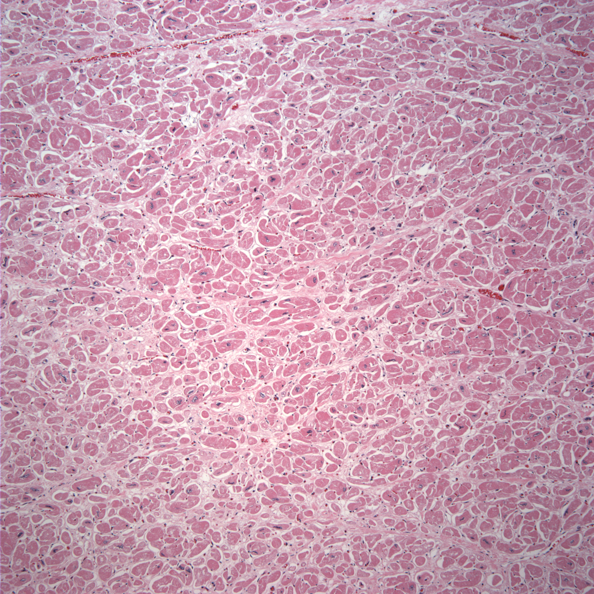
Case 1, image 2: There is extensive myocardial fibrosis in this case. Often, there are hypertrophied myocytes with enlarged boxcar-like nuclei.
)
Case 1, image 3: A closer look.
)
BACKGROUND
In hypertensive heart disease, often, the left ventricle is concentrically hypertrophied. Microscopically, there may be diffuse interstitial fibrosis.
Last updated: 2013-10-18
For questions, comments or feedback on this case: editor@surgpath4u.com