
Case 1, image 1: This was a 58 year old man who died of a ruptured myocardial infarction due to atherosclerotic cardiovascular disease.
)
Case 1, image 2: Blood is dissecting through myocardium. Some granulation tissue can be seen on the left.
)
Case 1, image 3: There is coagulative necrosis, hypereosiniophilia and loss of striations in the dying myocytes.
)
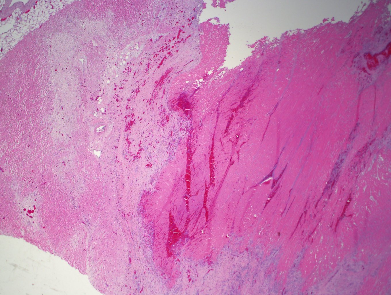
Case 1, image 4: Blood can be seen dissecting through granulation tissue, which indicates a healing infarct.
)
Case 2, image 1: This 78 year old man died of a ruptured myocardial infarction due to hypertensive and atherosclerotic cardiovascular disease.
)
Myocardial rupture occurs as a consequence of myocardial infarction (heart attack). The most common rupture site is left ventricular free wall. Free-wall rupture usually is most frequent three to eight days after a myocardial infarction when coagulative necrosis, neutrophilic infiltration, and lysis of myocardial connective tissue have weekend the infracted myocardium (injured heart muscle).