
IMAGE DESCRIPTIONS
Case 1, image 1: This was a 38 year old man who died of liver failure due to hepatitis C infection. Sections of the basal ganglia demonstrate Alzheimer type II astrocytes with cleared out nuclei.
)
Case 1, image 2: Another image of cleared out astrocytic nuclei that typify Alzheimer type II astrocytes.
)
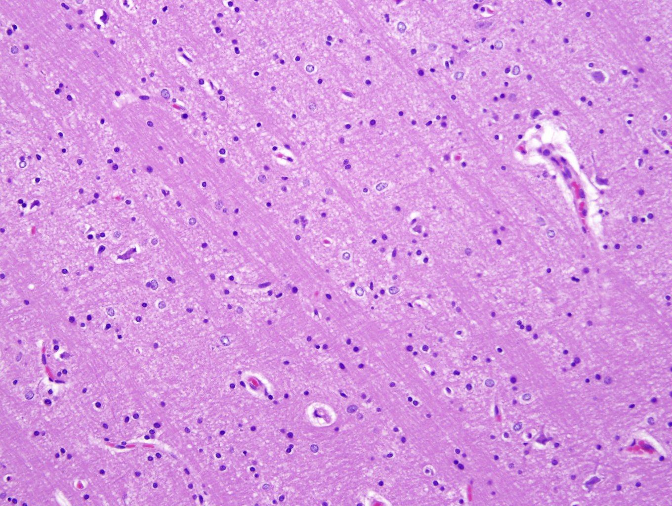
Case 1, image 3: A closer look at the swollen astrocytes.
)
BACKGROUND
Alzheimer type II astrocytes are indicative of hepatic encephalopathy and hyperammonemia. The basal ganglia is a good place to look for these reactive astrocytes, however, they can also be seen throughout the brain.
Last updated: 2013-10-15
For questions, comments or feedback on this case: editor@surgpath4u.com