
Case 1, image 1: This was a 72 year old woman who died of a subdural hematoma due to a blunt force injury of the head. She fell at home. This image demonstrates a fairly recent clot. Not shown was an area of old hemorrhage with granulation tissue.
)
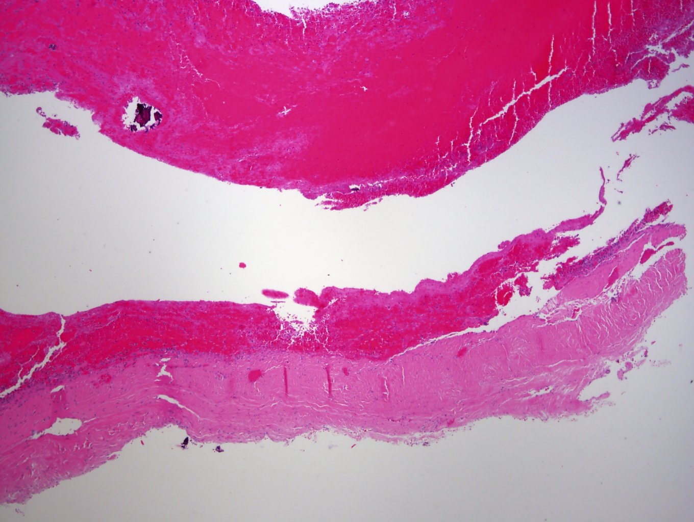
Case 1, image 2: There is some early fibroblastic activity at the clot-dura interface, which suggests that it has been 3-4 days since the hematoma occurred.
)
Case 2, image 1: This was a 5 month old infant who died of nonaccidental blunt trauma.
)
Case 2, image 2: Note that this is a well-formed neomembrane with bands of collagen on top of the dura mater, which suggests that at least several weeks (or more likely a month) has passed since the subdural hemorrhage occurred.
)
Case 2, image 3: There are vessels that form within the neomembrane.
)
Case 2, image 4: There was new hemorrhage within the dura in this child alongside the well-formed neomembrane. This indicates that new injury had occurred in addition to prior injury.
)
The formation and resolution of subdural hemorrhage follows a stepwise process and if sampled carefully, a forensic pathologist can cautiously give an approximate date of hemorrhage and in some instances, various ages of healing can suggest repeated trauma.
Cummings PM, Trelka DP, Springer KM (2011). Atlas of Forensic Histopathology. Cambridge, UK. Cambridge University Press.