
Case 1, image 1: This was a 23 year old woman who died of complications of anoxic encephalopathy secondary to multiple drug intoxication. Prior to death, she likely experienced a period of hypotension and hypoperfusion.
)
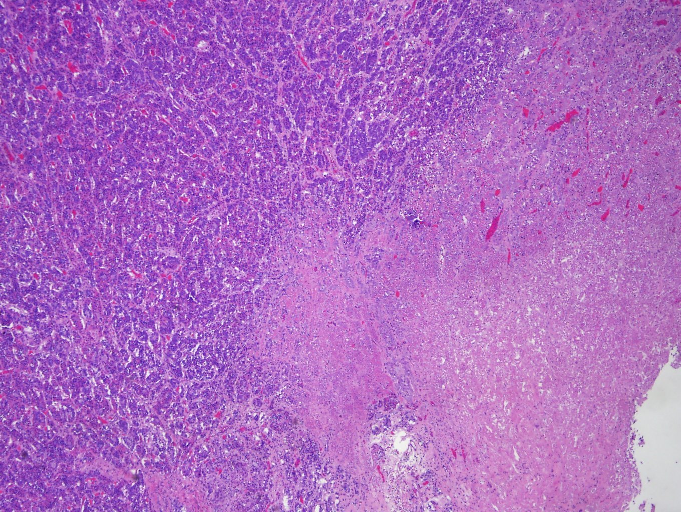
Case 1, image 2: Sections of the pitutiary show regional necrosis within the adenohypophysis.
)
Case 1, image 3: A closer look at infarcted glandular tissue.
)
Pituitary apoplexy describes a condition in which the pituitary gland becomes infarcted or hemorrhagic. Usually, it is an acute expansion of a pre-existing pituitary adenoma, however, the infarction or hemorrhage may occur in a nonadenomatous pituitary gland.
The cause of pituitary apoplexy may be systemic hypotension (i.e. sepsis) that causes hypoperfusion and infarction. Hemorrhage may occur as reperfusion after infarct or due to systemic anticoagulation, overstimulation of the pituitary by exogenous administration of ACTH, or rupture of blood vessels i.e. from tumor growth (Zink, 2005).
In pregnant women, the pituitary gland enlarges and the blood loss of childbirth can lead to hypoperfusion and infarction (Sheehan syndrome).
Zink E. Recognizing and Intervening in Pituitary Apoplexy. Topics in Advanced Practice Nursing eJournal. 2005;5(4). Available at http://www.medscape.com/viewarticle/518323_5