System: Forensic Pathology: Brain: : Multiple sclerosis

System: Forensic Pathology: Brain: : Multiple sclerosis

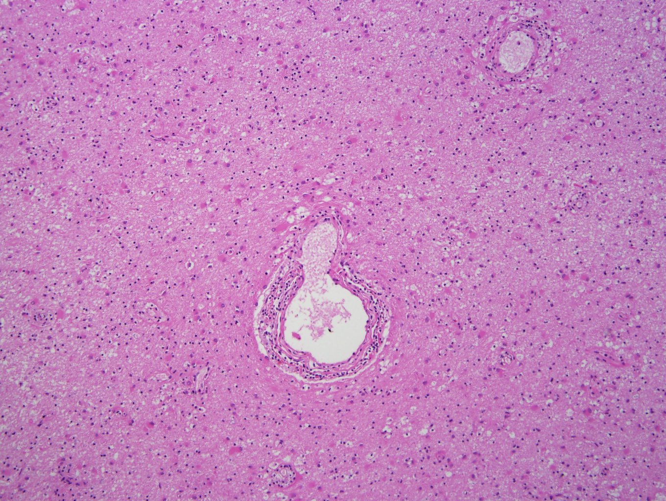
Case 1, image 1: This was a 37 year old man who died from suicidal ingestion of diphenydramine. His past medical history included multiple sclerosis and depression. There were active plaques with perivascular lymphocytes and evolving gliosis.
Case 1, image 2: A closer look at the perivascular lymphocytes of an acute MS plaque.
Case 1, image 3: Gross image of plaques.
Multiple sclerosis is characterized by focal areas of demyelination (plaques) predominantly in the white matter, although plaques may also be found within the cortex, thalamus and basal ganglia. The most common sites are the optic nerves and optic chiasm, periventricular and periaqueductal regions, floor of the fourth ventricle, and subpial regions of the spinal cord. Histologically, there are three stages: inflammation, myelin breakdown and gliosis.
)
)
)
)
)