
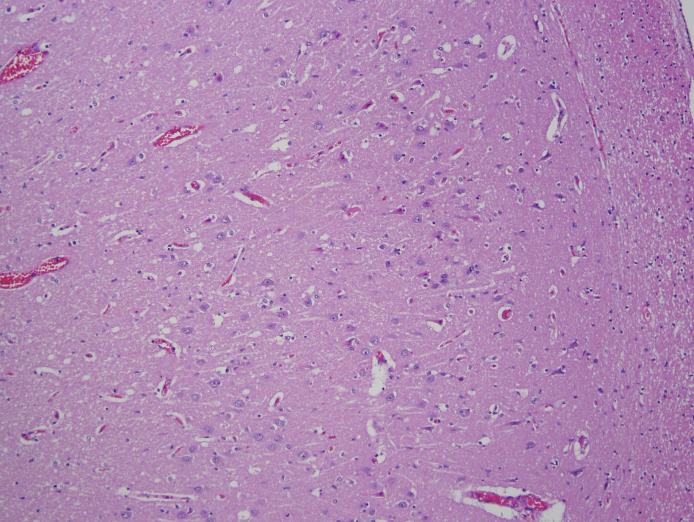
Case 1, image 1: This is a 55 year old woman who was found unresponsive in her jail cell in ventricular fibrillation. She was resuscitated and brought to the hospital where she developed anoxic encephaopathy. This is a section of her hippocampus.
)
Case 1, image 2: The neurons are not merely dark, but red.
)
Case 1, image 3: Hypereosinophilic cytoplasm and a degenerating nucleus.
)
The so-called red neurons are indicative of hypoxic change.
Of note, a forensic pathologist also trained in neuropathology stated that you may see these red neurons adjacent to areas of cerebral trauma. Physically injured neurons may also turn red (Forensic Pathology Blog, 2013)
Forensic Pathology Blog, uploaded July 23rd, 2013. Available at http://forensicmd.blogspot.com/2013/07/time-to-get-back-to-work.html