
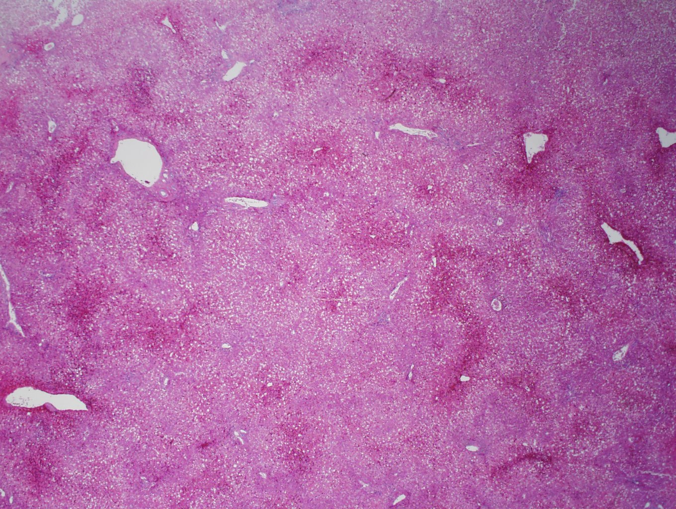
Case 1, image 1: This is a 61 year old man who became hypotensive due to sepsis. He became septic after his bowel infarcted (translocation of bacteria) due to his atherosclerotic blood vessels in his bowel. He also took a large amount of acetaminophen, presumably for this discomfort. At low power view of the liver, a pattern emerges.
)
Case 1, image 2: There is congestion and necrosis of hepatocytes predominantly around the venules. In this instance, both hypotension and acetaminophen toxicity contributed to centrilobular necrosis.
)
Case 1, image 3: The hepatocytes around the portal triads are mostly spared. Macrovesicular steatosis is fairly prominent, not surprising as this decedent consumed a lot of alcohol.
)
Case 2, image 1: This was a 57 year old woman who died of chronic lung disease. In this instance, acetaminophen was not detected and the centrilobular necrosis likely occured due to a period of decreased blood flow prior to death.
)
Case 2, image 2: The hepatocytes around the portal triads are spared, as you can see.
)
Case 2, image 3: Necrosis of hepatocytes around the central veins.
)
Case 3, image 1: This 40-year-old man died of acetaminophen (Tylenol) toxicity.
)
Case 3, image 2: There is a background of autolysis, however, one can still discern a pattern of pericentral hepatocyte necrosis
)
Case 3, image 3: The necrosis extends to zone 3 where a few, relatively intact, vacuolated hepatocytes are adjacent to portal tracts.
)
Centrilobular necrosis is most commonly seen in hypotension or acetaminophen toxicity.