System: Soft Tissue: Lipomatous: Neoplastic: Liposarcoma, Dedifferentiated with IMT-like Features

System: Soft Tissue: Lipomatous: Neoplastic: Liposarcoma, Dedifferentiated with IMT-like Features

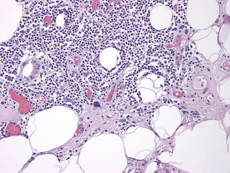
inflammatory well differentiated LS area Image
another areas of well differentiated LS Image
a spindled rather bland dediff area Image
a more cellular area Image
some focal atypia Image
another case with myxoid Image
ganglion like Image
atypical mits Image
cellular area Image
another case that is hypocellular Image
more of this Image
whorled areas Image
SMA on left, desmin on right Image
MDM2 Image
)
)
)
)
)
)
)
)
)
)
)
)
)
)