
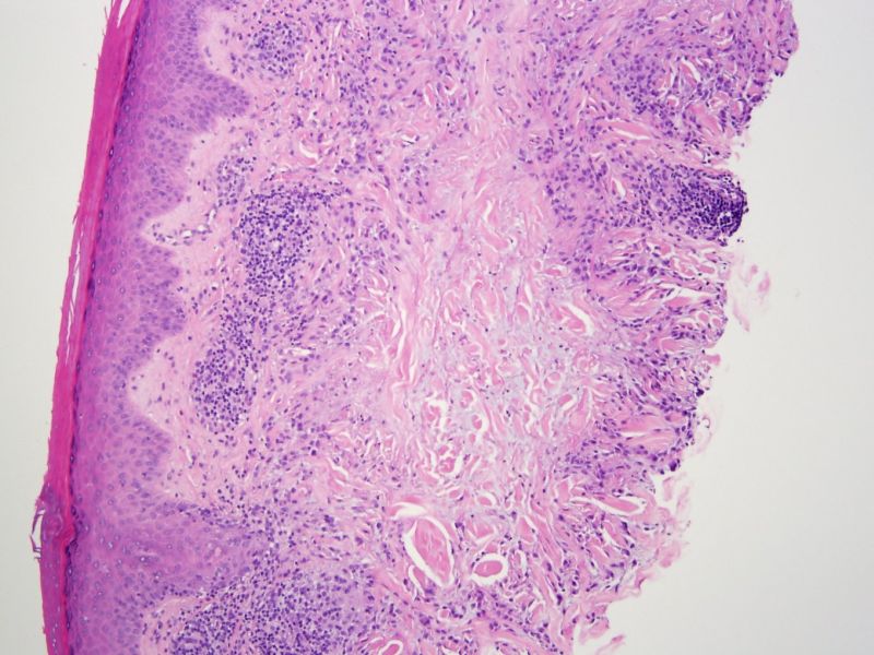
Granuloma annulare is characterized by a palisading granuloma composed of histiocytes and giant cells around a core of mucin and degenerating connective tissue (necrobiosis). The overlying epidermis is normal.
)
Histiocytes and multinucleated giant cells palisade around the core of degenerative connective tissue (necrobiotic material) and mucin.
)
A perivascular lymphocytic infiltrate and scattered eosinophils (not seen here) can be common. Neutrophils may also be present, but plasma cells are uncommon.
)
Granuloma annulare is an idiopathic annular papule or plaque that appear on the distal extremities of children and young adults (hands, feet, elbows) although older adults may be affected as well . There is central clearing of these flesh-colored or red raised plaques, thus, giving them an annular (ring-like) appearance.
The differential diagnosis includes necrobiosis lipoidica (granulomas with degenerating collagen and minimal mucin, presence of plasma cells) and rheumatoid nodules (fibrin in the center of the granuloma and no mucin). So pay attention to what is in the center of the granuloma.
It often has a ringed appearance to the plaques, or it occurs as small groups of papules. Patients are otherwise healty.
Usually no treatment is necessary, but steroids or cryotherapy can be applied for some cases of isolated GA. For the generalized variant of GA,isotretinoin or phototherapy has been used.
It often resolves on its own but may recur in the same or different sites.
Busam KJ. Dermatopathology: Foundations in Diagnostic Pathology 1st Ed. Philadelphia, PA: Elsevier; 2010: 51-2.
Rapini RP.Practical Dermatopathology. Philadelphia, PA: Elsevier; 2005: 99-100.