
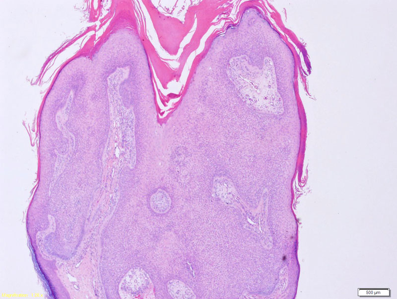
Tricholemmoma is a small circumscribed tumor located in the epidermis and extending down as broad nests to the upper reticular dermis. The periphery of the tumor demonstrate pallisading of basaloid cells resembling the outer root sheath of a hair follicle. The lobules are usually surrounded by a thick basement membrane. Keratinization and squamous eddies may be seen (Busam, Rapini). The surface often contains hyperkeratosis and sometimes verrucous change.
)
It is composed of lobules of cuboidal cells with light pink or clear cytoplasm (due to plentiful glyogen). These lobules connect to the epidermis or follicular infundibulum.
)
Usually presents as a solitary skin-colored papule on face of young adults. Most are solitary and sporadic. If multiple lesions are seen, it is a marker for Cowden syndrome.
80% of Cowden syndrome is caused by mutations in the PTEN tumor suppressor gene. Cowden syndrome or "multiple hamartomatous syndrome" is characterized by the development of multiple hamartomas on the skin (e.g. tricholemmomas, fibromas, acrochordons), mucosal membranes (oral mucosal papilomatosis) and GI tract. There is an increased risk of developing certain malignancies (e.g. breast, thryoid, endometrium) as well as neurological disability.
These lesions are benign.
→Multiple trichilemmomas may indicate Cowden disease.
→Histologic features include download pushing lobular growth of pale glycogenated keratinocytes. The basaloid cells palisade at the periphery of the lobules and are often surrounded by a thickened basement membrane.
Busam KJ. Dermatopathology: Foundations in Diagnostic Pathology 1st Ed. Philadelphia, PA: Elsevier; 2010: 386-7.
Rapini RP. Practical Dermatopathology. Philadelphia, PA: Elsevier; 2005: 290-2.