System: Skin: Lymphoid: Neoplastic: Sezary Syndrome

System: Skin: Lymphoid: Neoplastic: Sezary Syndrome

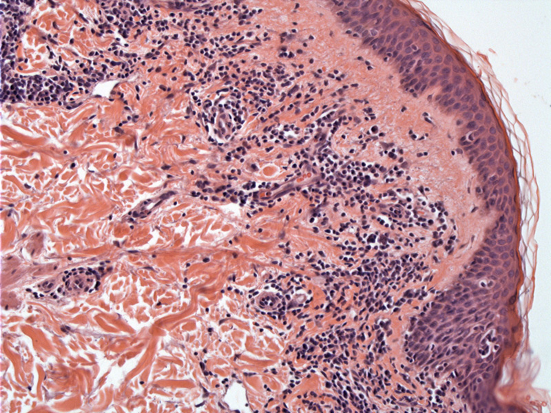
Case 1: The epidermis shows features consistent with mycosis fungoides, with a lichenoid infiltrate and epidermotropism of atypical lymphocytes.
At higher power, the atypical lymphocytes can be seen lining the basal layer as well as in the epidermis, creating punched out spaces.
Sezary syndrome is considered a more advanced form (leukemic phase) of mycosis fungoides (MF), occurring in approximately 5% of MF cases. Patients with Sezary syndrome generally have greater than 1000 Sezary cells per cubic mm in the peripheral blood, often accompanied by generalized exfoliative erythroderma and lymphoadenopathy (Rapini, Pinter-Brown).
On peripheral blood smear, the atypical T lymphocytes exhibit a characterisitic cerebriform nuclei. The T-cell rearrangement can be demonstrated by molecular or cytogenetic techniques. There is an increase of CD4+ (T-helper cells) and a loss of CD8+ (cytotoxic T cells) such that the ratio of CD4/CD8 is greater than 10. Aberrant loss of the pan-T cell marker CD7 has been thought to be helpful (Pinter-Brown).
→Sezary syndrome is considered an advanced phase of mycosis fungoides, the prototypical cutaneous T-cell lymphoma.
• Lymphoid : Mycosis Fungoides
Pinter-Brown LC. Cutaneous T-Cell Lymphoma Overview of CTCL. eMedicine. Last updated on May 17th 2011. Available at: emedicine.medscape.com/article/209091-overview
Rapini RP.Practical Dermatopathology. Philadelphia, PA: Elsevier; 2005: 309-310.
)
)